

Understanding the nerve innervation of the eye muscles is crucial for diagnosing and treating various ophthalmological conditions. These tiny but mighty muscles are responsible for controlling eye movements, allowing us to track objects, maintain focus, and coordinate our vision. This post delves into the specifics of which cranial nerves control which eye muscles.
The movement of each eye is controlled by six extraocular muscles: the superior rectus, inferior rectus, medial rectus, lateral rectus, superior oblique, and inferior oblique. Each of these muscles receives its nerve supply from a specific cranial nerve. The oculomotor nerve (cranial nerve III) is the primary driver, innervating four of these six muscles: the superior rectus (responsible for upward gaze), the inferior rectus (downward gaze), the medial rectus (adduction, moving the eye towards the nose), and the inferior oblique (elevation, abduction, and external rotation). It also innervates the levator palpebrae superioris, which elevates the upper eyelid.
The trochlear nerve (cranial nerve IV) innervates only one muscle: the superior oblique. This muscle is responsible for depression, abduction, and internal rotation of the eye. Because of its trajectory and how it inserts into the eye, its action is complex and essential for activities like reading and looking downwards.
Finally, the abducens nerve (cranial nerve VI) controls the lateral rectus muscle, which is responsible for abduction of the eye (moving the eye away from the nose). Problems with the abducens nerve often result in an inability to abduct the affected eye, leading to diplopia (double vision) due to misalignment.
A dysfunction in any of these cranial nerves can result in a variety of symptoms, including double vision (diplopia), drooping eyelids (ptosis), and abnormal eye movements. Understanding the precise nerve supplying each muscle allows clinicians to pinpoint the location of the nerve damage and determine the underlying cause.
Frequently Asked Questions (FAQ)
- Which nerve controls the lateral rectus muscle?
- The lateral rectus muscle is controlled by the abducens nerve (cranial nerve VI).
- What are the symptoms of oculomotor nerve palsy?
- Symptoms can include drooping eyelid (ptosis), dilated pupil, and difficulty moving the eye up, down, or inward.
- Which muscle does the trochlear nerve control?
- The trochlear nerve (cranial nerve IV) controls the superior oblique muscle.
- What happens if the abducens nerve is damaged?
- Damage to the abducens nerve can cause an inability to abduct the eye (move it outward), leading to double vision.
If you are searching about What Are the Parts of the Eye Diagram and Anatomy Explained Human eye – Wikiwand you’ve visit to the right place. We have 15 Pics about Introduction To The Eye Pasco Vision Clinic, What Are the Parts of the Eye Diagram and Anatomy Explained, Human eye – Wikiwand like ESA – Eye-catching space technology restoring sight Structure of the Eye MCAT Psychology MedSchoolCoach What Are the Parts of the Eye Diagram and Anatomy Explained Introduction To The Eye Pasco Vision Clinic. Here you go:
What Are The Parts Of The Eye Diagram And Anatomy Explained

Image description: Eye Image, Beautiful Eye Image, 5760×3840, 15459 Anatomy of the Eye Gallery of Photos
DIAGRAM Diagram Of The Eye – MYDIAGRAM.ONLINE Understanding The Human Eye Anatomy And Function
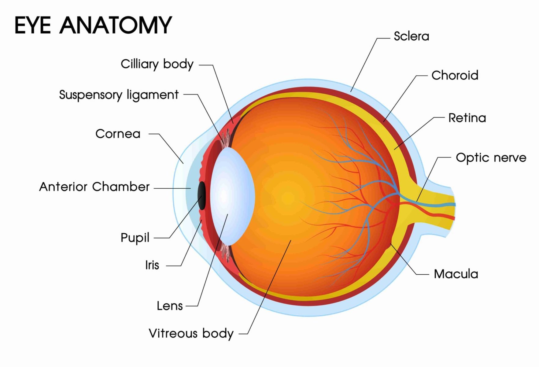
Image description: DIAGRAM Diagram Of The Eye – MYDIAGRAM.ONLINE Eye Anatomy Concise Medical Knowledge
Eye Anatomy Concise Medical Knowledge

Image description: Structure of the Eye MCAT Psychology MedSchoolCoach Human eye – Wikiwand Understanding the Human Eye Anatomy and Function Eye
ESA – Eye-catching Space Technology Restoring Sight

Image description: DIAGRAM Diagram Of The Eye – MYDIAGRAM.ONLINE A workout for your eyes Human eye – Wikipedia
ESA – Eye-catching Space Technology Restoring Sight

Image description: Introduction To The Eye Pasco Vision Clinic Human eye – Wikipedia
A Workout For Your Eyes Anatomical Structure Of The Eye. Source ClevelandClinic, 2022

Image description: Eye Anatomical structure of the eye. Source ClevelandClinic, 2022 What Are the Parts of the Eye Diagram and Anatomy Explained DIAGRAM Diagram Of The Eye – MYDIAGRAM.ONLINE
Anatomy Of The Eye Gallery Of Photos

Image description: Structure of the Eye MCAT Psychology MedSchoolCoach A workout for your eyes Eye Image, Beautiful Eye Image, 5760×3840, 15459
ESA – Eye-catching Space Technology Restoring Sight
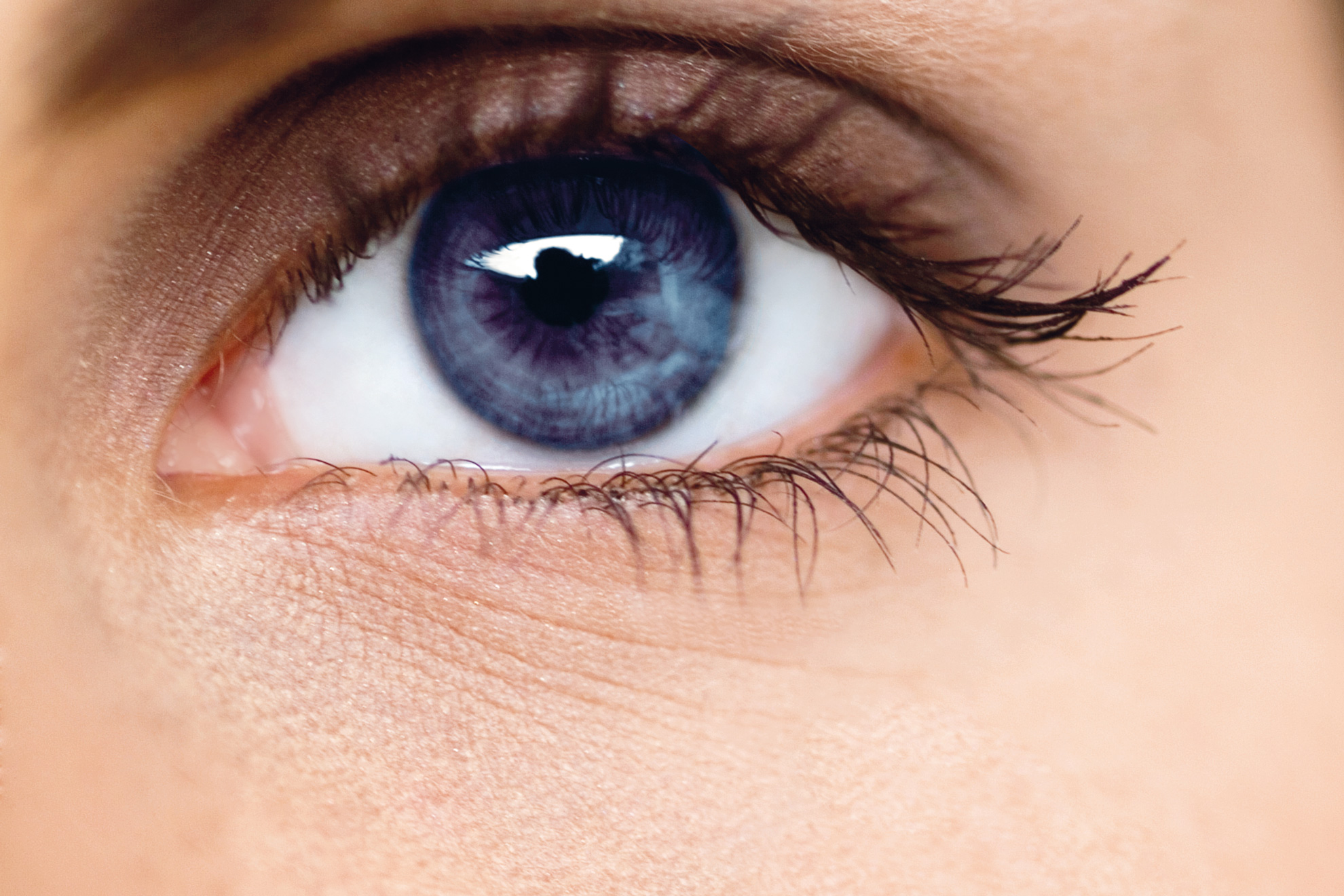
Image description: A workout for your eyes DIAGRAM Diagram Of The Eye – MYDIAGRAM.ONLINE Introduction To The Eye Pasco Vision Clinic
Eye A Workout For Your Eyes
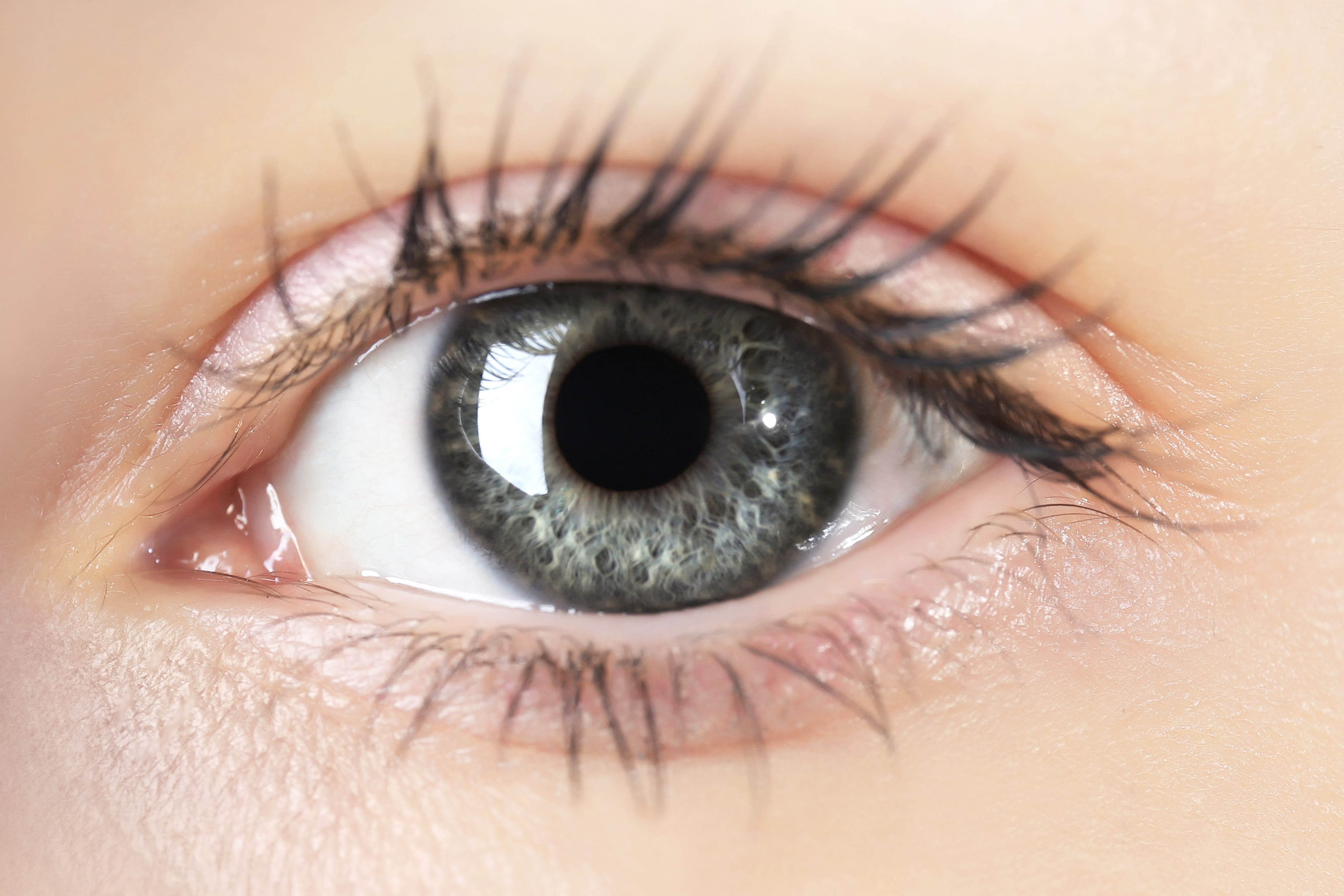
Image description: Anatomy of the Eye Gallery of Photos Human eye – Wikiwand What Are the Parts of the Eye Diagram and Anatomy Explained
Introduction To The Eye Pasco Vision Clinic Human Eye – Wikiwand
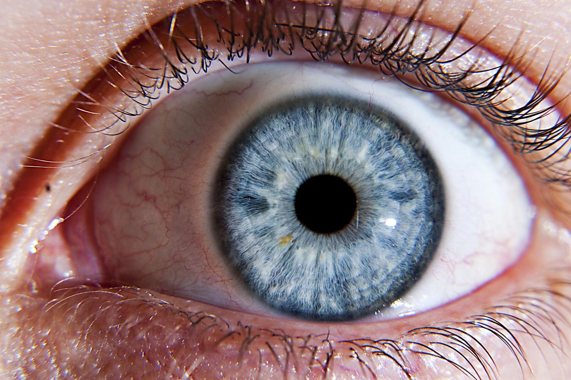
Image description: Anatomy of the Eye Gallery of Photos Understanding the Human Eye Anatomy and Function Anatomical structure of the eye. Source ClevelandClinic, 2022 Eye Definition, How does it Work, Anatomy, and Functions
ESA – Eye-catching Space Technology Restoring Sight What Are The Parts Of The Eye Diagram And Anatomy Explained
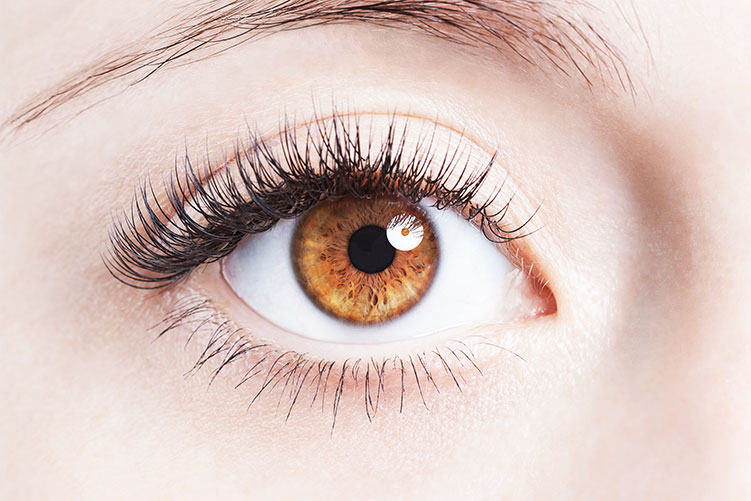
Image description: Introduction To The Eye Pasco Vision Clinic Structure of the Eye MCAT Psychology MedSchoolCoach
Eye Anatomy Concise Medical Knowledge Anatomical Structure Of The Eye. Source ClevelandClinic, 2022
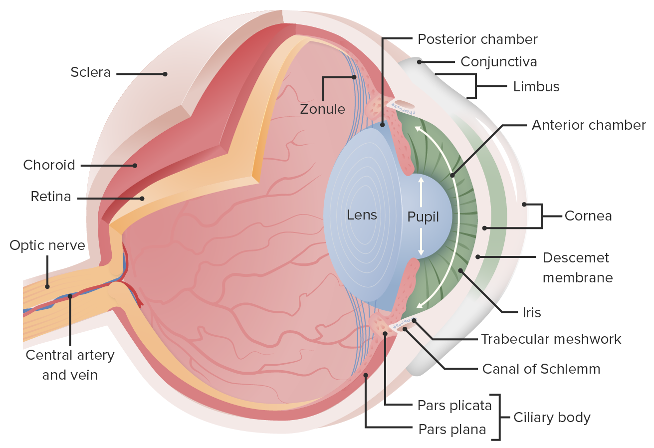
Image description: Eye Anatomy Concise Medical Knowledge Human eye – Wikiwand A workout for your eyes
Human Eye – Wikipedia
:max_bytes(150000):strip_icc()/GettyImages-553394069-59a34f61d963ac001178667d.jpg)
Image description: A workout for your eyes Structure of the Eye MCAT Psychology MedSchoolCoach Human eye – Wikipedia Anatomical structure of the eye. Source ClevelandClinic, 2022
Human Eye – Wikipedia Anatomy Of The Eye Gallery Of Photos
![[DIAGRAM] Diagram Of The Eye - MYDIAGRAM.ONLINE](https://fm.cnbc.com/applications/cnbc.com/resources/img/editorial/2018/08/14/105395023-1534234590550gettyimages-746122853.1910x1000.jpeg)
Image description: Eye Anatomy Concise Medical Knowledge DIAGRAM Diagram Of The Eye – MYDIAGRAM.ONLINE
Eye Definition, How Does It Work, Anatomy, And Functions
/GettyImages-1176128997-50151a347f6645e98e944a7b55fba595.jpg)
Image description: ESA – Eye-catching space technology restoring sight A workout for your eyes Eye Image, Beautiful Eye Image, 5760×3840, 15459
Images featured are entirely for illustrative intentions only. We don’t keep any outside media on our servers. Visual content is linked directly from royalty-free sources used for non-commercial use only. Downloads are delivered straight from the source distributors. For any intellectual property issues or content removal demands, please get in touch with our moderator using the Contact page.


