

Understanding the intricate movements of our eyes requires knowledge of the nerves that control the extraocular muscles. These muscles, situated around the outside of each eyeball, are responsible for directing our gaze in various directions. Let’s delve into the fascinating world of eye muscle nerve supply.
The control of eye movements isn’t as simple as one nerve per eye. In fact, three cranial nerves work in concert to provide innervation to the six extraocular muscles of each eye: the oculomotor nerve (CN III), the trochlear nerve (CN IV), and the abducens nerve (CN VI). Each nerve has a specific role in coordinating precise and synchronized movements, ensuring binocular vision and preventing double vision (diplopia).
The oculomotor nerve (CN III) is the workhorse of the group, innervating four of the six extraocular muscles. These muscles are the superior rectus (responsible for upward movement), inferior rectus (downward movement), medial rectus (adduction, moving the eye towards the nose), and inferior oblique (extorsion, elevation when adducted, and abduction when depressed). Additionally, the oculomotor nerve also controls the levator palpebrae superioris, which elevates the upper eyelid, and carries parasympathetic fibers to the pupillary constrictor muscle and the ciliary muscle, responsible for pupillary constriction and accommodation (focusing for near vision), respectively.
The trochlear nerve (CN IV) innervates only one muscle: the superior oblique. This muscle has a unique anatomical course, passing through a cartilaginous pulley called the trochlea before inserting onto the eyeball. The superior oblique is primarily responsible for intorsion (rotating the top of the eye towards the nose), depression when adducted, and abduction when elevated. It plays a crucial role in looking down and outwards.
The abducens nerve (CN VI) also innervates a single muscle: the lateral rectus. As its name suggests, the lateral rectus is responsible for abduction, moving the eye away from the nose. This allows us to look sideways.
Damage to any of these nerves can result in characteristic patterns of eye movement deficits, leading to diplopia and difficulty coordinating eye movements. A thorough neurological examination is essential in diagnosing and managing these conditions.
Frequently Asked Questions (FAQ)
- Which nerve controls the superior rectus muscle?
- The oculomotor nerve (CN III) controls the superior rectus muscle.
- What happens if the abducens nerve is damaged?
- Damage to the abducens nerve (CN VI) results in an inability to abduct the affected eye. The eye will be pulled inward due to the unopposed action of the medial rectus, leading to horizontal diplopia.
- Why are there three nerves controlling eye movements?
- Three nerves are required to precisely coordinate the complex movements of the six extraocular muscles in each eye, allowing for smooth pursuit, saccades (rapid eye movements), and vergence (converging or diverging the eyes). This intricate system ensures binocular vision and depth perception.
- What is diplopia?
- Diplopia, or double vision, occurs when the images from each eye are not properly aligned and fused by the brain. It can be a symptom of damage to the nerves or muscles that control eye movements.
If you are searching about Structure of the Eye MCAT Psychology MedSchoolCoach Human Eyes Images you’ve visit to the right web. We have 15 Images about Anatomical structure of the eye. Source ClevelandClinic, 2022, Eye, Eyeball Anatomy Pictures like Anatomical structure of the eye. Source ClevelandClinic, 2022 Human Eyes Images Evolution of the eye New Scientist Human eye – Wikiwand. Here you go:
Eyeball Anatomy Pictures

Image description: Human eye – Wikiwand Eye Definition, How does it Work, Anatomy, and Functions DIAGRAM Diagram Of The Eye – MYDIAGRAM.ONLINE Structure of the Eye MCAT Psychology MedSchoolCoach
3 Anatomy Surrounding The Eye – OpticianWorks Online Optician Training Diagram Of Parts Of Eye Eyes How They Work, Anatomy Commo

Image description: Anatomical structure of the eye. Source ClevelandClinic, 2022 Human Eyes Images Eye Introduction To The Eye Pasco Vision Clinic
What Are The Parts Of The Eye Diagram And Anatomy Explained
/GettyImages-1176128997-50151a347f6645e98e944a7b55fba595.jpg)
Image description: Human eye – Wikiwand Eye
Anatomy Of The Eye Gallery Of Photos

Image description: Human eye – Wikiwand Diagram Of Parts Of Eye Eyes How They Work, Anatomy Commo DIAGRAM Diagram Of The Eye – MYDIAGRAM.ONLINE
Eye Definition, How Does It Work, Anatomy, And Functions Understanding The Human Eye Anatomy And Function
![[DIAGRAM] Diagram Of The Eye - MYDIAGRAM.ONLINE](https://fm.cnbc.com/applications/cnbc.com/resources/img/editorial/2018/08/14/105395023-1534234590550gettyimages-746122853.1910x1000.jpeg)
Image description: Human Eyes Images Human eye – Wikiwand What Are the Parts of the Eye Diagram and Anatomy Explained Eye Definition, How does it Work, Anatomy, and Functions
What Are The Parts Of The Eye Diagram And Anatomy Explained

Image description: 3 Anatomy Surrounding the Eye – OpticianWorks Online Optician Training Diagram Of Parts Of Eye Eyes How They Work, Anatomy Commo
Eyeball Anatomy Pictures Anatomical Structure Of The Eye. Source ClevelandClinic, 2022

Image description: Diagram Of Parts Of Eye Eyes How They Work, Anatomy Commo Evolution of the eye New Scientist Human eye – Wikiwand
What Are The Parts Of The Eye Diagram And Anatomy Explained

Image description: What Are the Parts of the Eye Diagram and Anatomy Explained Eye Definition, How does it Work, Anatomy, and Functions Diagram Of Parts Of Eye Eyes How They Work, Anatomy Commo Eyeball Anatomy Pictures
Diagram Of Parts Of Eye Eyes How They Work, Anatomy Commo

Image description: Understanding the Human Eye Anatomy and Function Eye What Are the Parts of the Eye Diagram and Anatomy Explained DIAGRAM Diagram Of The Eye – MYDIAGRAM.ONLINE
Eye Definition, How Does It Work, Anatomy, And Functions 3 Anatomy Surrounding The Eye – OpticianWorks Online Optician Training

Image description: Human Eyes Images Diagram Of Parts Of Eye Eyes How They Work, Anatomy Commo
DIAGRAM Diagram Of The Eye – MYDIAGRAM.ONLINE

Image description: Human Eyes Images DIAGRAM Diagram Of The Eye – MYDIAGRAM.ONLINE Human eye – Wikiwand
Evolution Of The Eye New Scientist
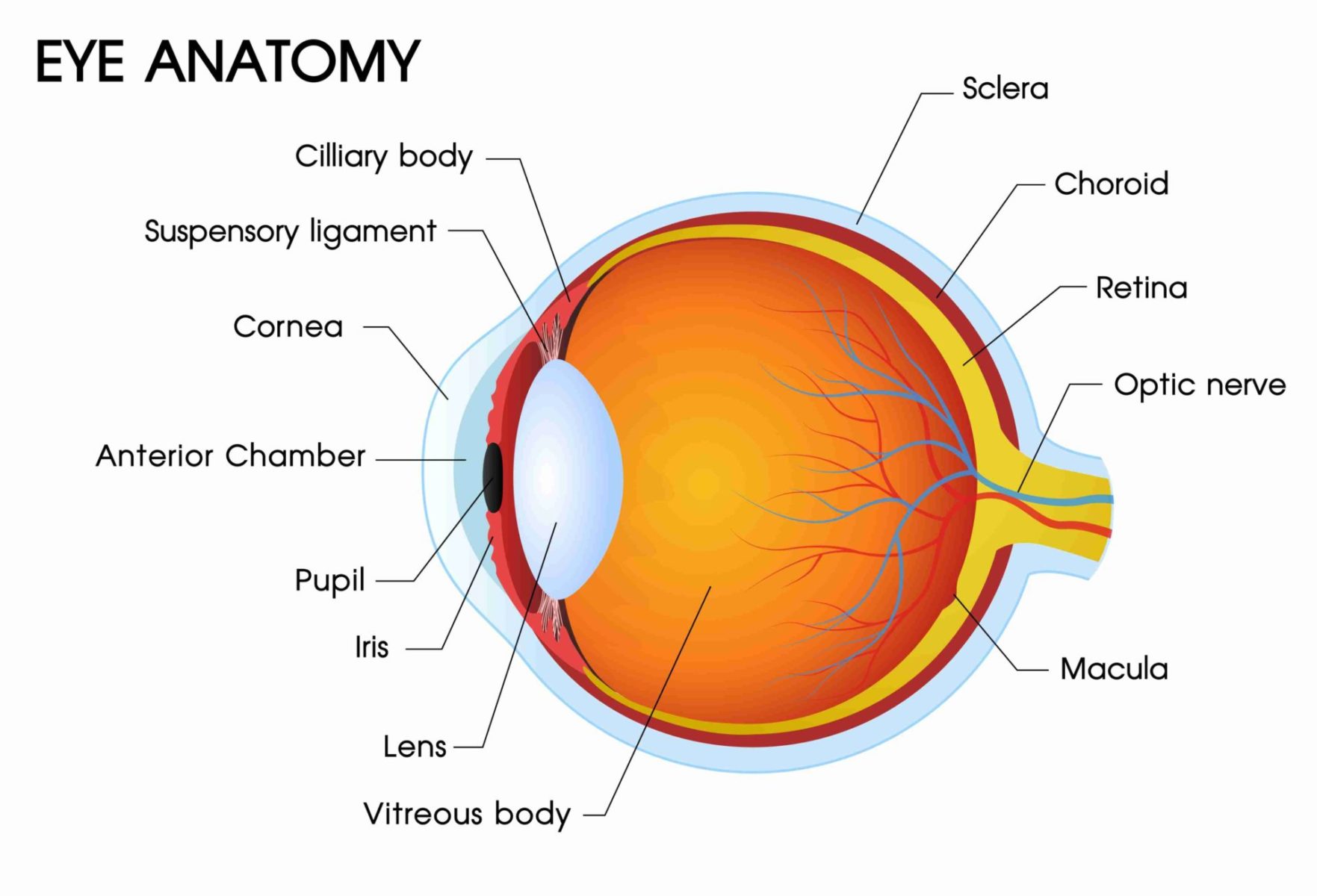
Image description: Understanding the Human Eye Anatomy and Function Anatomical structure of the eye. Source ClevelandClinic, 2022 Anatomy of the Eye Gallery of Photos
3 Anatomy Surrounding The Eye – OpticianWorks Online Optician Training
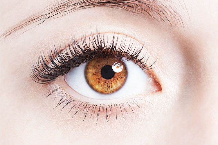
Image description: Anatomical structure of the eye. Source ClevelandClinic, 2022 Evolution of the eye New Scientist Anatomy of the Eye Gallery of Photos
Anatomy Of The Eye Gallery Of Photos Eye Definition, How Does It Work, Anatomy, And Functions
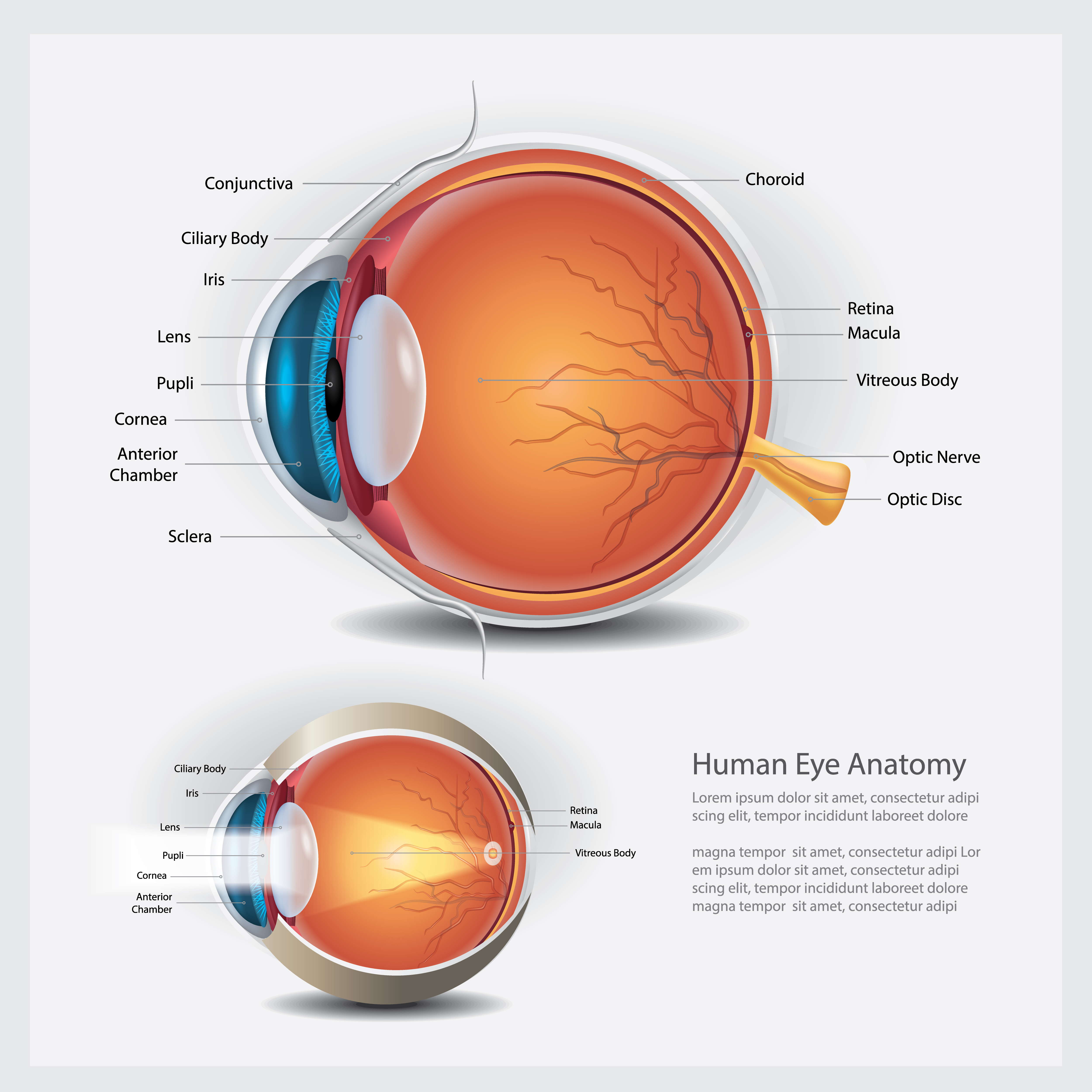
Image description: Anatomical structure of the eye. Source ClevelandClinic, 2022 Human eye – Wikiwand Eyeball Anatomy Pictures Eye
Diagram Of Parts Of Eye Eyes How They Work, Anatomy Commo Structure Of The Eye MCAT Psychology MedSchoolCoach
:max_bytes(150000):strip_icc()/GettyImages-553394069-59a34f61d963ac001178667d.jpg)
Image description: Anatomy of the Eye Gallery of Photos DIAGRAM Diagram Of The Eye – MYDIAGRAM.ONLINE Evolution of the eye New Scientist Eyeball Anatomy Pictures
Images featured are purely for educational objectives only. We do not host any off-site media on our servers. Media is embedded straightforwardly from royalty-free sources intended for non-commercial use only. Downloads are offered straight from the official websites. For any copyright concerns or requests for removal, please communicate with our staff through Contact page.


