

Understanding the femoral nerve and the muscles it innervates is crucial for comprehending lower limb function and diagnosing related neurological conditions. This post will delve into the specifics of femoral nerve innervation, providing a clear explanation of which muscles receive their nerve supply from this important nerve.
The femoral nerve, originating from the lumbar plexus (L2-L4 spinal nerve roots), is the largest nerve in the lumbar plexus and a primary nerve responsible for the anterior thigh compartment’s motor and sensory functions. It descends through the psoas major muscle, emerging along its lateral border, and then travels beneath the inguinal ligament to enter the thigh. Once in the thigh, it splits into numerous branches that provide both motor and sensory innervation.
The primary motor function of the femoral nerve involves innervating the muscles responsible for knee extension and hip flexion. These muscles include the quadriceps femoris group: the rectus femoris, vastus lateralis, vastus medialis, and vastus intermedius. The rectus femoris contributes to both hip flexion and knee extension. The other three vastus muscles (lateralis, medialis, and intermedius) act solely on knee extension. Collectively, these muscles allow us to straighten our leg at the knee, essential for walking, running, and jumping. Furthermore, the femoral nerve also innervates the sartorius muscle, the longest muscle in the human body. The sartorius muscle facilitates hip flexion, abduction, and external rotation, as well as knee flexion and internal rotation. Finally, the pectineus muscle, responsible for hip flexion and adduction, receives partial innervation from the femoral nerve (it may also receive innervation from the obturator nerve).
In addition to its motor functions, the femoral nerve also provides sensory innervation to the anterior and medial thigh, as well as the medial side of the leg and foot via the saphenous nerve, a terminal cutaneous branch of the femoral nerve.
Frequently Asked Questions (FAQ)
- What spinal levels does the femoral nerve originate from? The femoral nerve originates from the L2, L3, and L4 spinal nerve roots.
- What is the main function of the muscles innervated by the femoral nerve? The primary function is knee extension, facilitated by the quadriceps femoris group, and hip flexion, contributed by the rectus femoris, sartorius, and pectineus.
- Which muscles are innervated by the femoral nerve? The quadriceps femoris (rectus femoris, vastus lateralis, vastus medialis, vastus intermedius), sartorius, and pectineus (partially) muscles.
- What is the saphenous nerve? The saphenous nerve is a terminal cutaneous branch of the femoral nerve that provides sensory innervation to the medial side of the leg and foot.
- What happens if the femoral nerve is damaged? Damage to the femoral nerve can result in weakness or paralysis of the quadriceps muscles, leading to difficulty extending the knee and problems with walking. Sensory loss can also occur in the anterior and medial thigh and medial leg.
If you are searching about Femoral triangle Anatomy Zone Femoral Artery at Don Kimber blog you’ve visit to the right web. We have 15 Pictures about The Femoral Nerve Entrapment And Anterior Thigh Pain, Femoral Artery Diagram, Femoral Femoral Bypass Jackie Kustan Medical Illustra – vrogue.co like Femoral Artery A Femoralis The Arteries Of The Lower Femoral Artery Supplies Muscles at James Browning blog Anthrpina Systimton tou Smatos Practica 2 Femur Head. Here it is:
Femoral Artery Medical School Stuff, Medical School Essentials, Basic Plaque In Femoral Artery At Clyde Rucker Blog

Image description: The Femoral Nerve Entrapment And Anterior Thigh Pain Femoral triangle Femoral Artery Diagram
Femoral Triangle Floor

Image description: Plaque In Femoral Artery at Clyde Rucker blog Anatomy Zone Femoral Artery at Don Kimber blog
Superficial Femoral Artery Diagram Femoral Nerve What Is It,
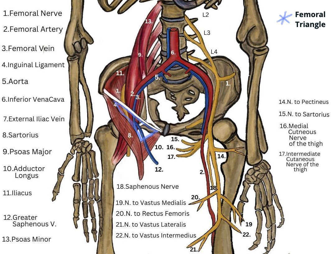
Image description: Anthrpina Systimton tou Smatos Practica 2 Plaque In Femoral Artery at Clyde Rucker blog The Femoral Nerve Entrapment And Anterior Thigh Pain Superficial Femoral Artery Anatomy
Superficial Femoral Artery Diagram Femoral Nerve What Is It,
Image description: Femoral Triangle Floor Femoral Artery Supplies Muscles at James Browning blog
Superficial Femoral Artery Diagram Femoral Nerve What Is It,
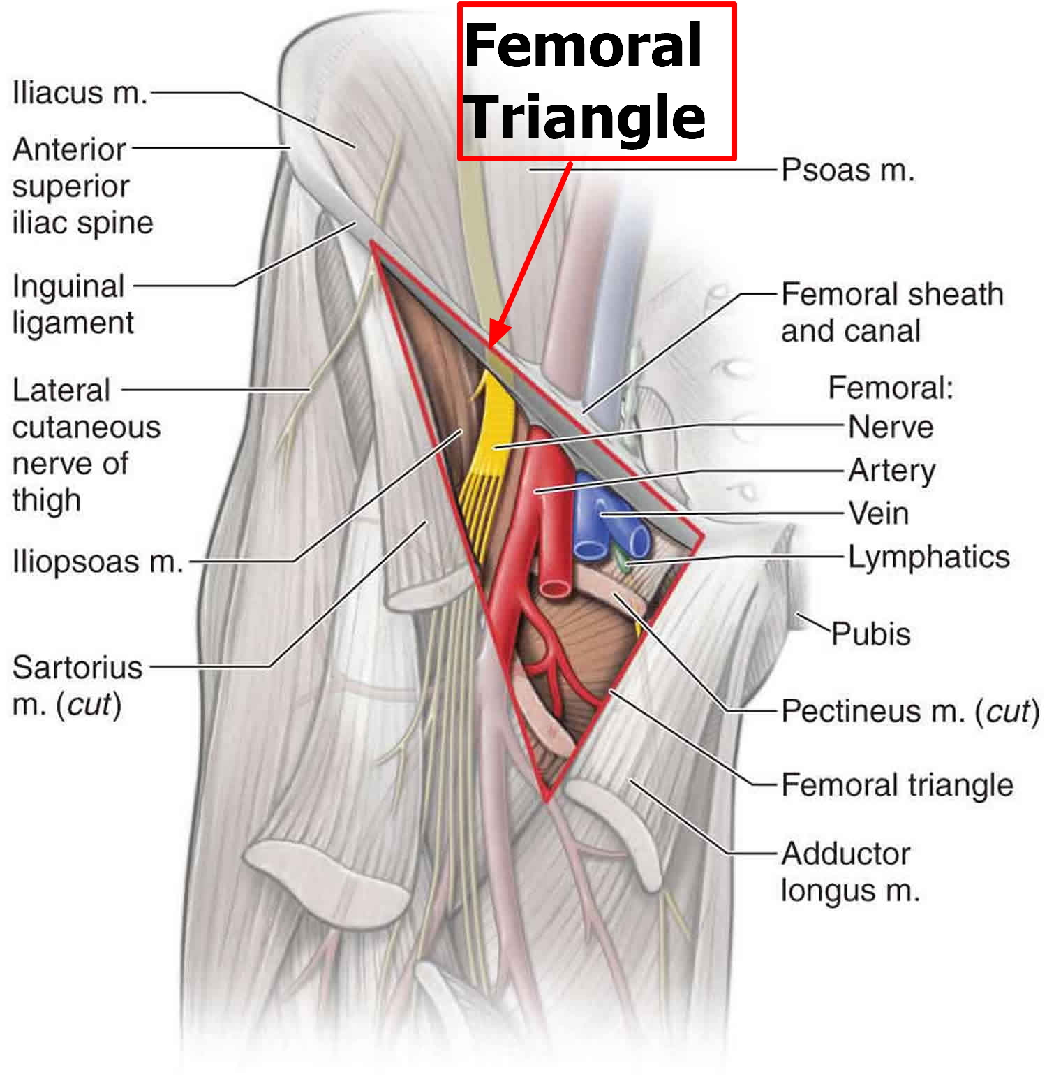
Image description: Femoral artery Medical school stuff, Medical school essentials, Basic Superficial Femoral Artery Diagram Femoral Nerve What Is It, Superficial Femoral Artery Anatomy
Femoral Artery A Femoralis The Arteries Of The Lower Superficial Femoral Artery Anatomy
Image description: Superficial Femoral Artery Anatomy Femur Head
Anthrpina Systimton Tou Smatos Practica 2
Image description: Femoral Triangle Floor Anthrpina Systimton tou Smatos Practica 2 Femoral artery Medical school stuff, Medical school essentials, Basic Femur Head
Femoral Femoral Bypass Jackie Kustan Medical Illustra – Vrogue.co
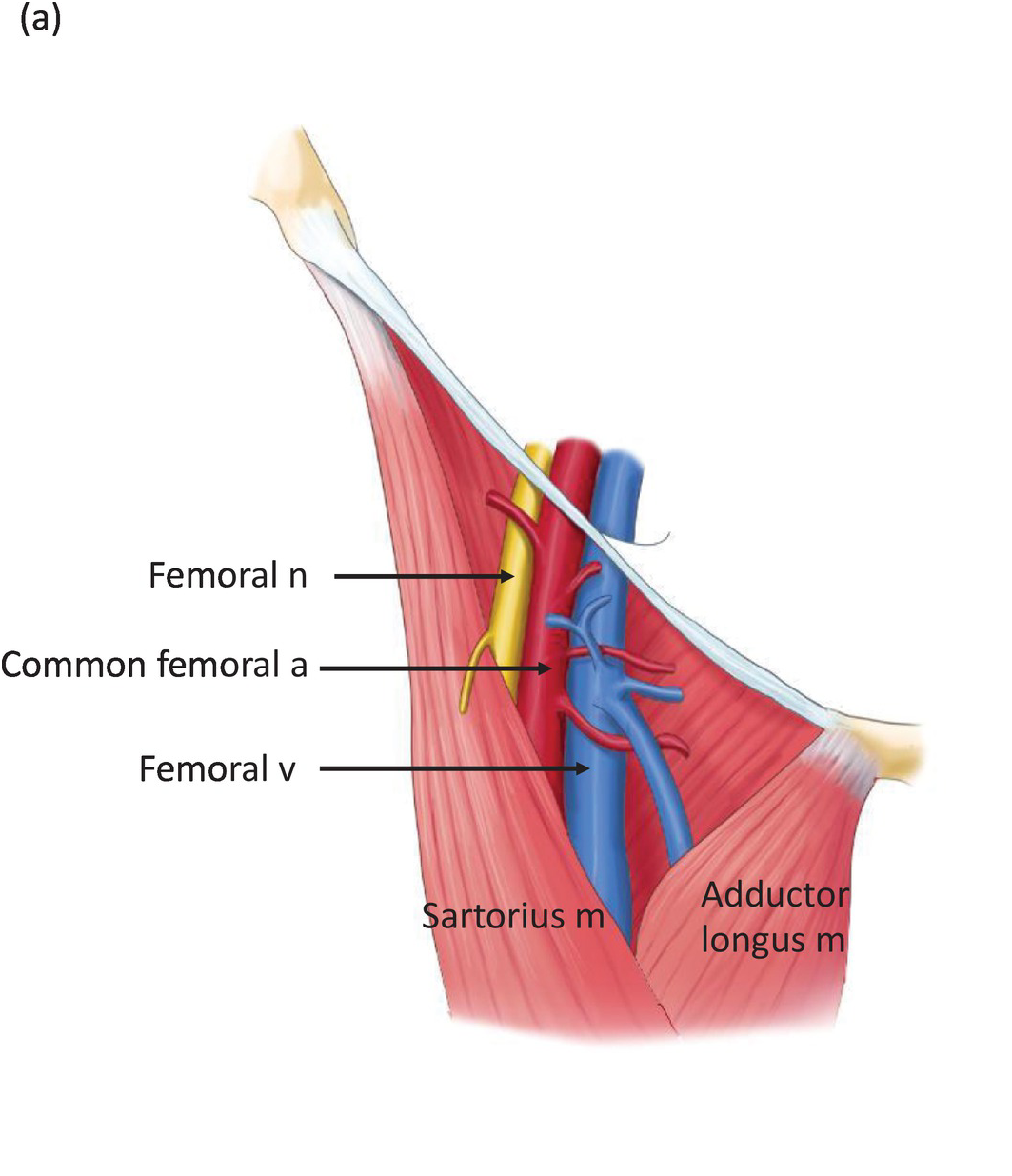
Image description: Radiopaedia – Drawing Contents and boundaries of the femoral triangle Femoral artery Medical school stuff, Medical school essentials, Basic Femoral triangle
Femoral Triangle
.jpeg)
Image description: Femoral artery Medical school stuff, Medical school essentials, Basic Anatomy Zone Femoral Artery at Don Kimber blog
Femur Head

Image description: Anatomy Zone Femoral Artery at Don Kimber blog Femoral Artery Diagram
Femur Head Femoral Artery Supplies Muscles At James Browning Blog

Image description: Radiopaedia – Drawing Contents and boundaries of the femoral triangle Femur Head
Femoral Artery Diagram Femoral Artery A Femoralis The Arteries Of The Lower
Image description: The Femoral Nerve Entrapment And Anterior Thigh Pain Femoral triangle
The Femoral Nerve Entrapment And Anterior Thigh Pain
Image description: Anatomy Zone Femoral Artery at Don Kimber blog Femoral Artery Supplies Muscles at James Browning blog
Radiopaedia – Drawing Contents And Boundaries Of The Femoral Triangle

Image description: Femur Head Anatomy Zone Femoral Artery at Don Kimber blog
Superficial Femoral Artery Diagram Femoral Nerve What Is It,
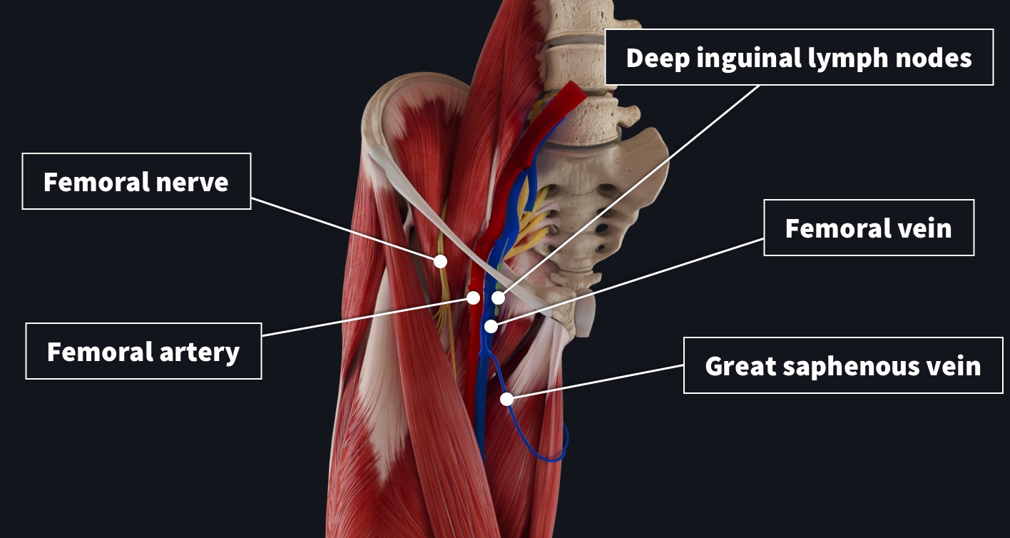
Image description: Femoral Triangle Floor Radiopaedia – Drawing Contents and boundaries of the femoral triangle Femoral Artery Diagram
Visuals shown here are purely for illustrative use only. Our servers do not store any third-party media on our system. The materials is embedded directly from royalty-free sources shared for non-commercial use only. Resources are served straight from the official providers. For any ownership inquiries or deletion requests, please communicate with our staff through our Contact page.


