

Understanding the iliopsoas muscle and potential issues can be complex. Thankfully, ultrasound imaging provides a non-invasive and readily accessible method to visualize this important muscle group. This post delves into the benefits, uses, and what to expect from an iliopsoas muscle ultrasound.
An iliopsoas muscle ultrasound is a diagnostic imaging technique that uses sound waves to create real-time images of the iliopsoas muscle. The iliopsoas, located deep within the abdomen and pelvis, is actually two muscles – the iliacus and the psoas major – that merge together and insert on the lesser trochanter of the femur. It’s a key hip flexor, crucial for walking, running, and maintaining posture. Because of its deep location, direct examination can be challenging. Ultrasound provides a valuable tool for assessing the muscle’s size, structure, and presence of any abnormalities. The procedure is relatively quick, painless, and doesn’t involve ionizing radiation, making it a safe option for most patients.
Iliopsoas muscle ultrasound is commonly used to evaluate various conditions affecting this muscle group. One frequent application is in diagnosing iliopsoas bursitis, an inflammation of the bursa (a fluid-filled sac that cushions tendons and bones) located near the muscle. The ultrasound can detect fluid accumulation within the bursa, confirming the diagnosis and helping to guide treatment. It’s also used to identify muscle strains or tears, which can occur due to overuse, trauma, or sudden movements. The ultrasound can reveal the extent of the injury and guide rehabilitation strategies. Another important use is in the assessment of iliopsoas abscesses, collections of pus within the muscle. Early detection of an abscess is crucial to prevent serious complications, and ultrasound can help guide drainage procedures. In children, iliopsoas muscle ultrasound is sometimes used to evaluate hip pain or a limp, particularly in cases of suspected septic arthritis or other inflammatory conditions.
During an iliopsoas muscle ultrasound, you’ll typically lie on your back while a trained sonographer applies a gel to the lower abdomen and hip area. This gel helps improve contact between the ultrasound probe and your skin. The sonographer will then move the probe across your skin, capturing images of the iliopsoas muscle in different planes. You might be asked to move your leg or hip during the examination to allow for better visualization of the muscle. The entire process usually takes between 15 and 30 minutes. The images are viewed in real-time by the sonographer, and a radiologist will typically review the images and provide a written report to your doctor. Your doctor will then discuss the findings with you and determine the appropriate course of treatment, if necessary.
Frequently Asked Questions (FAQ)
-
Is an iliopsoas muscle ultrasound painful?
No, the procedure is generally painless. You might feel some pressure from the ultrasound probe, but it shouldn’t be uncomfortable.
-
Do I need to prepare for the ultrasound?
In most cases, no special preparation is required. Your doctor might advise you to wear loose-fitting clothing for comfort.
-
How quickly will I get the results?
The radiologist’s report usually takes a few days to be finalized. Your doctor will then discuss the results with you.
-
What are the risks of an iliopsoas muscle ultrasound?
Ultrasound is a very safe imaging technique. There are no known risks associated with the procedure.
If you are searching about 17 Musculoskeletal Ultrasound Radiology Key Shoulder Ultrasound Radiology Key you’ve visit to the right place. We have 15 Images about Elbow Ultrasound Radiology Key, Shoulder Ultrasound Radiology Key, Ultrasound Radiology Key like Shoulder Ultrasound Clinical Gate Elbow Ultrasound Radiology Key. Read more:
Right Knee Of A 36-year-old Male Showing A Normal Medial Meniscus

Image description: Knee Ultrasound Radiology Key Elbow Ultrasound Radiology Key
Ultrasound Radiology Key Shoulder Ultrasound Radiology Key
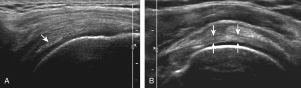
Image description: Muscle Injury and Sequelae – Clinical Tree Ultrasound Radiology Key
Right Knee Of A 36-year-old Male Showing A Normal Medial Meniscus Shoulder Ultrasound Radiology Key

Image description: Shoulder Ultrasound Radiology Key 17 Musculoskeletal Ultrasound Radiology Key Right knee of a 36-year-old male showing a normal medial meniscus
Hip Ultrasound Radiology Key Elbow Ultrasound Radiology Key

Image description: Hip Ultrasound Radiology Key Shoulder Ultrasound Radiology Key
Right Knee Of A 36-year-old Male Showing A Normal Medial Meniscus

Image description: Ultrasound Radiology Key Knee Ultrasound Radiology Key Hip Ultrasound Radiology Key
Hip Ultrasound Radiology Key
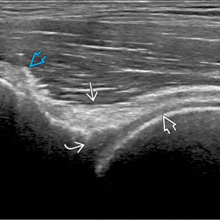
Image description: Muscle Injury and Sequelae – Clinical Tree Elbow Ultrasound Radiology Key Shoulder Ultrasound Radiology Key Shoulder Ultrasound Radiology Key
Muscle Injury And Sequelae – Clinical Tree

Image description: Ultrasound Radiology Key Quadriceps Tendon Ultrasound Representative ultrasound images of the rectus femoris muscle in a Elbow Ultrasound Radiology Key
Shoulder Ultrasound Radiology Key

Image description: Hip Ultrasound Radiology Key Representative ultrasound images of the rectus femoris muscle in a Right knee of a 36-year-old male showing a normal medial meniscus Ultrasound Radiology Key
17 Musculoskeletal Ultrasound Radiology Key Hip Ultrasound Radiology Key

Image description: Knee Ultrasound Radiology Key Ultrasound-Guided Iliopsoas Bursal Injections for Management of Ultrasound Radiology Key Shoulder Ultrasound Clinical Gate
Knee Ultrasound Radiology Key Ultrasound Radiology Key
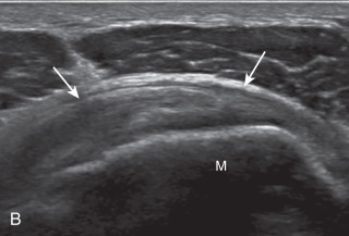
Image description: Shoulder Ultrasound Radiology Key Hip Ultrasound Radiology Key 17 Musculoskeletal Ultrasound Radiology Key Quadriceps Tendon Ultrasound
Muscle Injury And Sequelae – Clinical Tree Knee Ultrasound Radiology Key

Image description: Shoulder Ultrasound Radiology Key Representative ultrasound images of the rectus femoris muscle in a Knee Ultrasound Radiology Key
Muscle Injury And Sequelae – Clinical Tree

Image description: Shoulder Ultrasound Clinical Gate Shoulder Ultrasound Radiology Key
Shoulder Ultrasound Clinical Gate

Image description: Ultrasound images of anterior tibial muscle group, showing line of Representative ultrasound images of the rectus femoris muscle in a Knee Ultrasound Radiology Key 17 Musculoskeletal Ultrasound Radiology Key
Muscle Injury And Sequelae – Clinical Tree Quadriceps Tendon Ultrasound
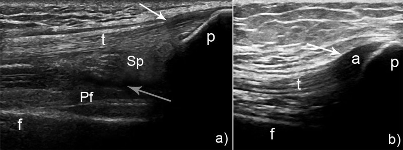
Image description: Ultrasound images of anterior tibial muscle group, showing line of Knee Ultrasound Radiology Key
Quadriceps Tendon Ultrasound

Image description: Shoulder Ultrasound Radiology Key Right knee of a 36-year-old male showing a normal medial meniscus
Images featured are solely for reference intentions only. We avoid storing any remote media on our platform. Visual assets is streamed automatically from royalty-free sources provided for informative use only. Assets are fetched straight from the legitimate platforms. For any copyright concerns or content removal demands, please contact our support team by using Contact page.


