

Ever wondered how your muscles actually move? It all comes down to a fascinating process occurring at the cellular level! Let’s dive into the amazing world of muscle cell contraction with a brief explanation that could be easily represented with an animation.
Muscle cell contraction is driven by the sliding filament theory, a captivating dance between two key protein filaments: actin (thin filaments) and myosin (thick filaments). Imagine myosin as tiny, molecular “rowers” ready to pull on actin “ropes”. These myosin rowers have “heads” that can bind to specific sites on the actin filaments. This binding is only possible when calcium ions are present. When a nerve impulse arrives at the muscle cell, it triggers the release of calcium ions from the sarcoplasmic reticulum, a network of internal membranes within the muscle cell. The presence of calcium then allows the myosin heads to attach to the actin. Once attached, the myosin heads pivot, pulling the actin filaments towards the center of the sarcomere (the basic unit of muscle contraction). This “power stroke” shortens the sarcomere. ATP (adenosine triphosphate), the cell’s energy currency, is crucial for this process. ATP binds to the myosin head, causing it to detach from the actin. The myosin head then re-energizes by breaking down the ATP, preparing it for another power stroke. This cycle repeats rapidly, causing the actin and myosin filaments to slide past each other, resulting in muscle contraction. When the nerve impulse stops, calcium ions are pumped back into the sarcoplasmic reticulum. Without calcium, the myosin heads can no longer bind to actin, and the muscle relaxes. The filaments slide back to their original positions, lengthening the sarcomere. Think of it as a microscopic tug-of-war, powered by ATP and controlled by calcium!
An animation of this process would clearly show the myosin heads grabbing, pulling, and releasing the actin filaments in a continuous, cyclical motion. It would also visualize the crucial role of calcium ions in initiating the process and ATP in powering it. A well-designed animation can make this complex process much easier to understand than just reading about it. Visualizing the sliding filament theory brings it to life!
Frequently Asked Questions
What are actin and myosin?
Actin and myosin are the two main protein filaments responsible for muscle contraction. Actin forms the thin filaments, while myosin forms the thick filaments.
What role does calcium play in muscle contraction?
Calcium ions are essential for muscle contraction. They bind to proteins on the actin filament, allowing myosin heads to attach and initiate the sliding filament mechanism.
What is ATP, and why is it important?
ATP (adenosine triphosphate) is the energy currency of the cell. It provides the energy needed for the myosin heads to detach from the actin filaments and reset for another power stroke.
What is the sarcomere?
The sarcomere is the basic functional unit of muscle contraction. It’s the segment between two Z lines in muscle tissue.
If you are looking for Chapter 3 The Muscular System Muscle discovery, aging and memory, plus other stories Mirage News you’ve came to the right page. We have 15 Pictures about Skeletal Muscle – Anatomy, Function, Structure, Types, Parts of a Skeletal Muscle, Shapes of skeletal muscles with various muscular types outline diagram like Muscle discovery, aging and memory, plus other stories Mirage News Skeletal Muscle Tissue Labeled Diagram What Muscles Do Pull-Ups Work. Here you go:
Quadriceps Muscle Diagram

Image description: Parts of a Skeletal Muscle Skeletal Muscle Tissue Labeled Diagram
Muscle Men On Cam
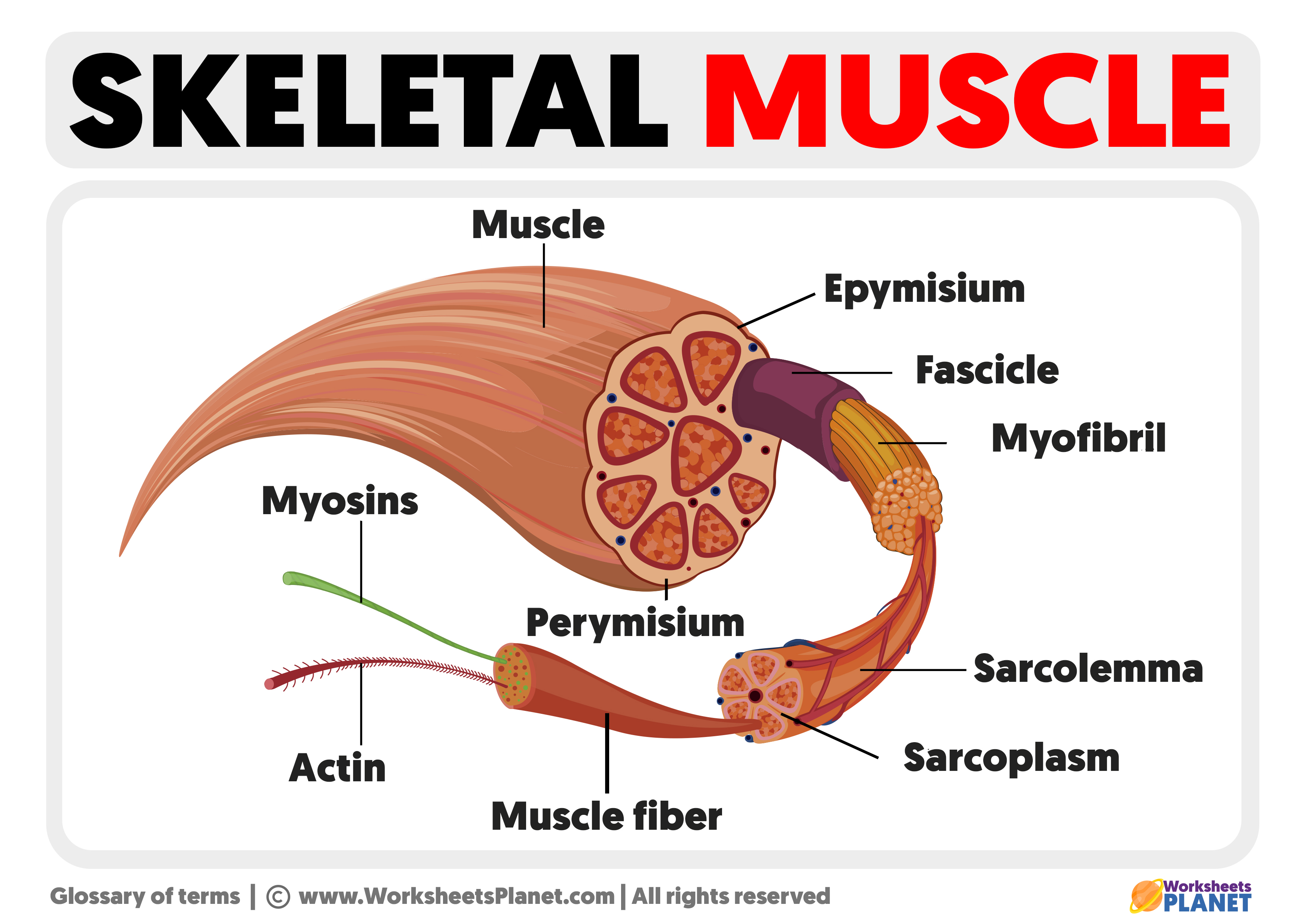
Image description: Parts of a Skeletal Muscle Skeletal muscle structure with anatomical inner layers outline diagram Arm Muscles Human Anatomy Image, Functions, Diseases and Treatments
Skeletal Muscle Structure With Anatomical Inner Layers Outline Diagram
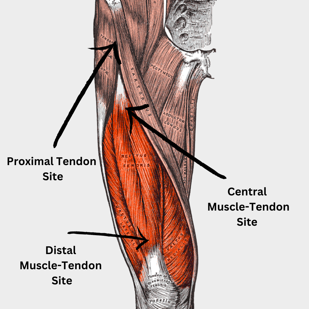
Image description: Skeletal Muscle Tissue Labeled Diagram What Muscles Do Pull-Ups Work Muscle discovery, aging and memory, plus other stories Mirage News
Muscle Structure Labeled, Illustrati Stock Image – Science Source Parts Of A Skeletal Muscle

Image description: Skeletal Muscle Tissue Labeled Diagram Shapes of skeletal muscles with various muscular types outline diagram
Arm Muscles Human Anatomy Image, Functions, Diseases And Treatments

Image description: Skeletal Muscle – Anatomy, Function, Structure, Types Arm Muscles Human Anatomy Image, Functions, Diseases and Treatments
Muscle Men On Cam Quadriceps Muscle Diagram

Image description: Skeletal Muscle Tissue Labeled Diagram Muscle Structure labeled, illustrati Stock Image – Science Source Shapes of skeletal muscles with various muscular types outline diagram Parts of a Skeletal Muscle
Parts Of A Skeletal Muscle
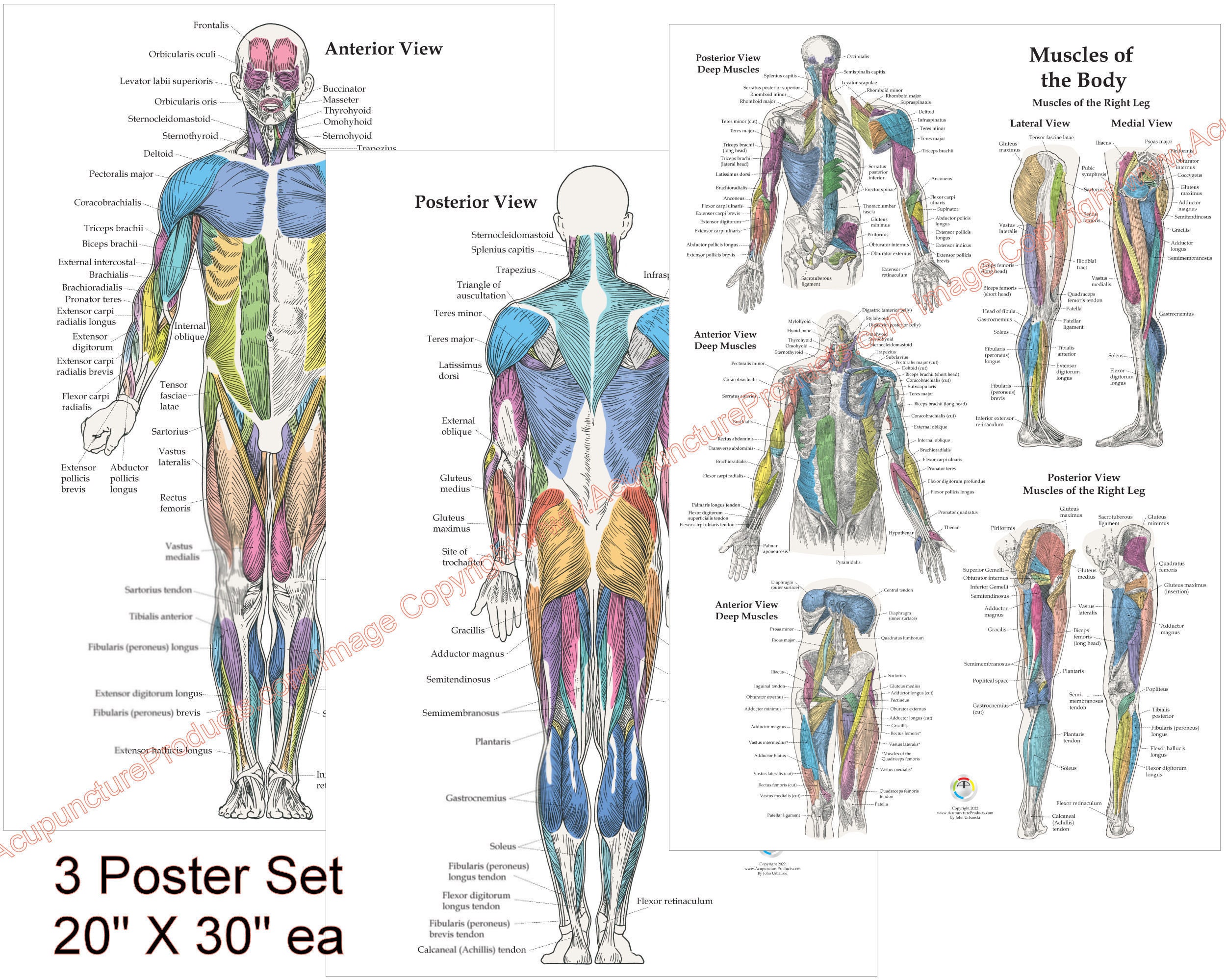
Image description: Skeletal muscle structure with anatomical inner layers outline diagram Chapter 3 The Muscular System Muscle Men On Cam What Muscles Do Pull-Ups Work
3 Human Muscle Anatomy Poster Anterior Posterior And Deep – Etsy Hong Kong

Image description: 3 Human Muscle Anatomy Poster Anterior Posterior and Deep – Etsy Hong Kong What Muscles Do Pull-Ups Work Chapter 3 The Muscular System
Download Athletic Shirtless Muscle Man Wallpaper Wallpapers.com Muscle Discovery, Aging And Memory, Plus Other Stories Mirage News
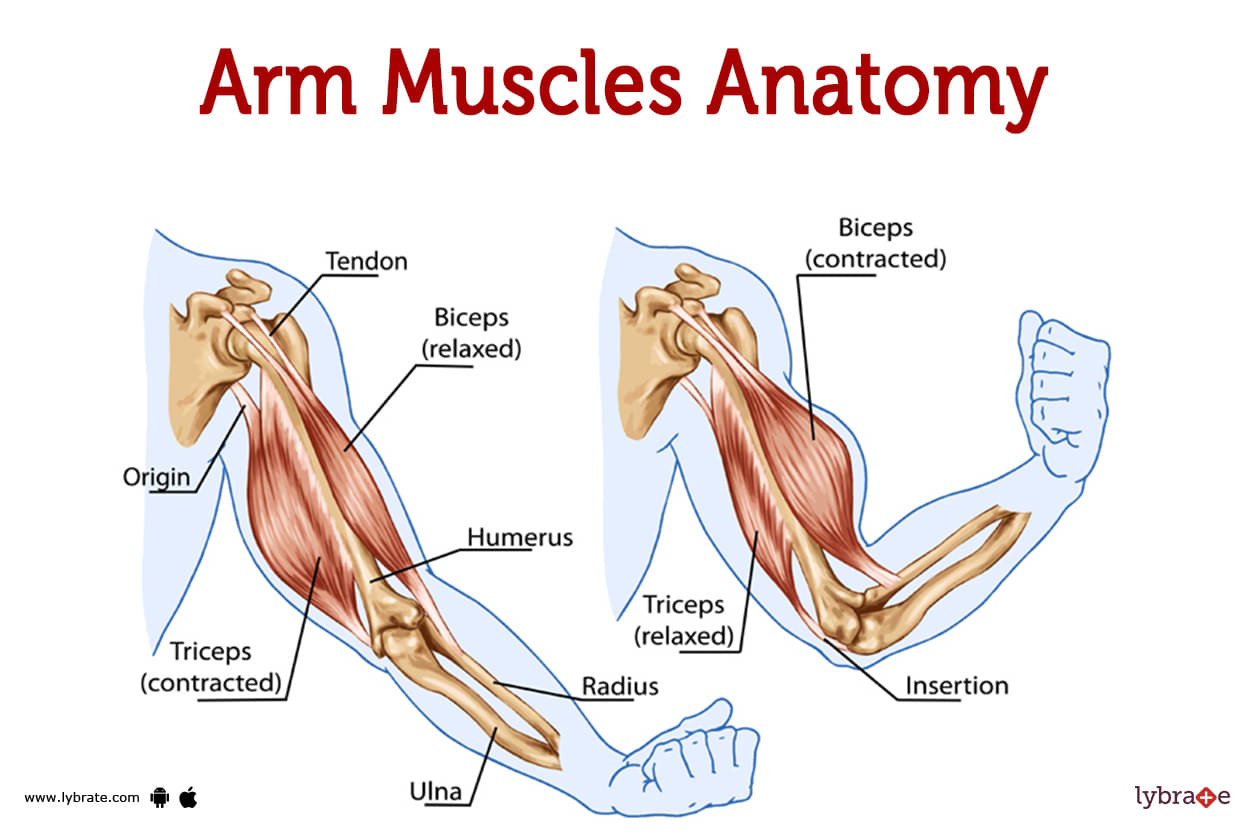
Image description: Parts of a Skeletal Muscle Muscle discovery, aging and memory, plus other stories Mirage News Anatomy Of The Upper Arm Arm Muscles Human Anatomy Image, Functions, Diseases and Treatments
Skeletal Muscle Structure With Anatomical Inner Layers Outline Diagram
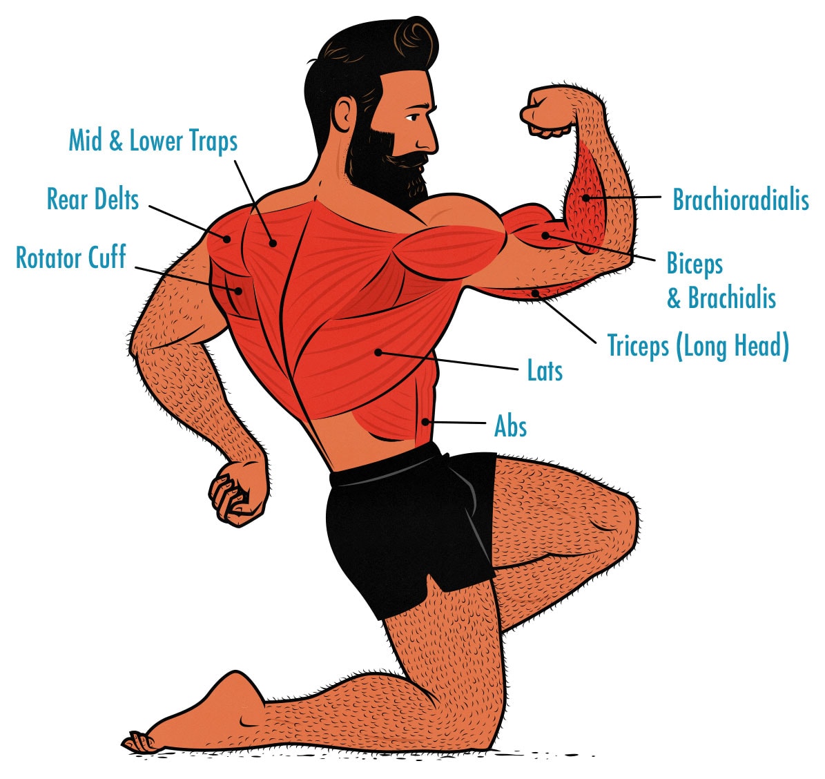
Image description: Parts of a Skeletal Muscle Muscle Men On Cam
Parts Of A Skeletal Muscle Chapter 3 The Muscular System

Image description: Muscle Structure labeled, illustrati Stock Image – Science Source Arm Muscles Human Anatomy Image, Functions, Diseases and Treatments
3 Human Muscle Anatomy Poster Anterior Posterior And Deep – Etsy Hong Kong Skeletal Muscle – Anatomy, Function, Structure, Types

Image description: 3 Human Muscle Anatomy Poster Anterior Posterior and Deep – Etsy Hong Kong What Muscles Do Pull-Ups Work Parts of a Skeletal Muscle
Chapter 3 The Muscular System
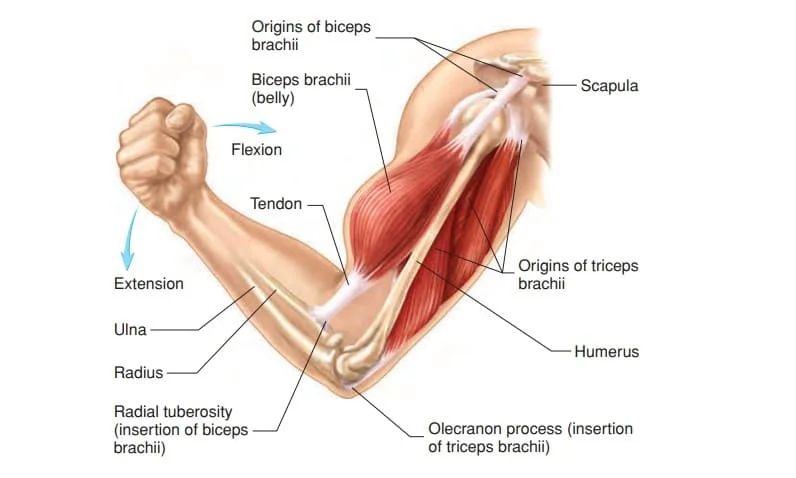
Image description: Shapes of skeletal muscles with various muscular types outline diagram 3 Human Muscle Anatomy Poster Anterior Posterior and Deep – Etsy Hong Kong Skeletal Muscle – Anatomy, Function, Structure, Types Download Athletic Shirtless Muscle Man Wallpaper Wallpapers.com
Arm Muscles Human Anatomy Image, Functions, Diseases And Treatments
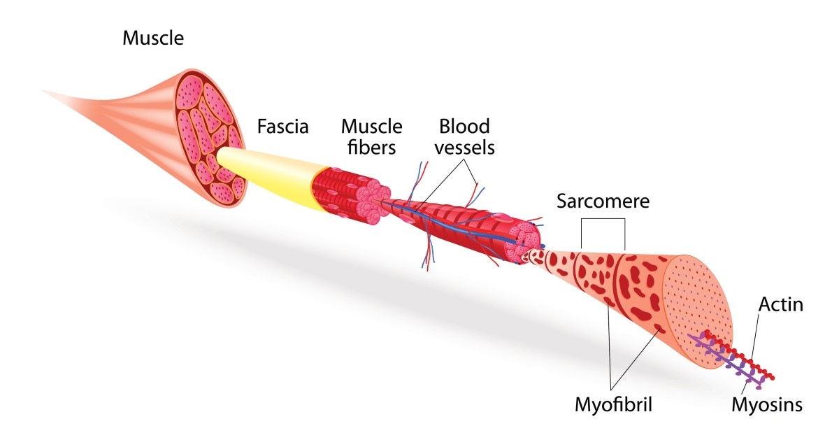
Image description: Arm Muscles Human Anatomy Image, Functions, Diseases and Treatments Muscle Structure labeled, illustrati Stock Image – Science Source Quadriceps Muscle Diagram
Muscle Structure Labeled, Illustrati Stock Image – Science Source

Image description: Muscle Men On Cam Download Athletic Shirtless Muscle Man Wallpaper Wallpapers.com
All images displayed are entirely for illustrative reasons only. We don’t keep any off-site media on our servers. Visual assets is integrated automatically from public domain sources meant for non-profit use only. Files are fetched straight from the official distributors. For any rights violations or takedown notices, please contact our moderator by using Contact page.


