

Understanding the muscles around the knee is crucial for anyone interested in fitness, physical therapy, or simply maintaining healthy mobility. This area is a complex intersection of muscles that control movement, provide stability, and protect the knee joint from injury. Let’s dive into the key muscles influencing the knee.
The knee joint, a hinge joint allowing flexion and extension (bending and straightening), relies heavily on surrounding muscles for its function and stability. The primary muscles responsible for knee extension (straightening the leg) are the quadriceps femoris, a group of four muscles located on the front of the thigh. These include the rectus femoris, vastus lateralis, vastus medialis, and vastus intermedius. The rectus femoris is unique as it also crosses the hip joint, contributing to hip flexion. All four quadriceps muscles converge and insert onto the tibial tuberosity via the patellar tendon, which encloses the patella (kneecap). Weakness in the quadriceps can contribute to knee instability and pain.
On the posterior (back) side of the thigh, the hamstring muscles are primarily responsible for knee flexion (bending the leg). This group consists of the biceps femoris, semitendinosus, and semimembranosus. These muscles originate from the ischial tuberosity (the “sit bone”) and insert below the knee. The biceps femoris inserts on the fibular head, while the semitendinosus and semimembranosus insert on the medial aspect of the tibia. The hamstrings also contribute to hip extension, acting as antagonists to the quadriceps during movements like walking and running.
Beyond the major movers, other muscles play a supporting role. The gastrocnemius, a calf muscle, crosses the knee joint and assists with knee flexion, although its primary function is plantarflexion of the foot (pointing the toes). Several smaller muscles, such as the sartorius, gracilis, and popliteus, also contribute to knee stability and fine motor control. The sartorius, originating at the hip and inserting on the medial tibia, assists with hip flexion, abduction, and external rotation, as well as knee flexion. The gracilis, located on the inner thigh, adducts the hip and assists with knee flexion. Finally, the popliteus, located behind the knee, assists with knee flexion and internal rotation of the tibia when the knee is flexed.
Understanding the anatomy and function of these muscles is vital for preventing injuries, optimizing performance, and rehabilitating after an injury. Strengthening these muscles, along with proper stretching and warm-up routines, can significantly reduce the risk of knee problems.
Frequently Asked Questions
Q: What happens if my quadriceps are weak?
A: Weak quadriceps can lead to knee instability, difficulty extending the knee, and increased risk of patellar tendinitis and other knee injuries.
Q: How can I strengthen my hamstrings?
A: Effective exercises for hamstring strengthening include hamstring curls, deadlifts, glute bridges, and Nordic hamstring exercises.
Q: What’s the best way to stretch the muscles around my knee?
A: Common stretches include quadriceps stretches, hamstring stretches (like touching your toes), and calf stretches. Hold each stretch for 20-30 seconds and repeat several times.
Q: Can tight muscles contribute to knee pain?
A: Yes, tight quadriceps, hamstrings, or calf muscles can restrict knee movement and contribute to pain and discomfort. Regular stretching is important.
If you are looking for Muscle Photo Muscular System Anatomy and Physiology – Nurseslabs you’ve visit to the right web. We have 15 Pics about Muscle Cell – Comparison Types of muscles, Tissue biology, Muscle tissue, Skeletal Muscle Cell Structure, Parts of a Skeletal Muscle like Parts of a Skeletal Muscle Muscle Anatomy Wallpapers – Top Free Muscle Anatomy Backgrounds. Here you go:
Muscular System Anatomy And Physiology – Nurseslabs Alphamale Teen Muscleman Flexing Huge Biceps Video
Image description: Muscle Anatomy Wallpapers – Top Free Muscle Anatomy Backgrounds Muscle Tissue – online presentation
Parts Of A Skeletal Muscle Skeletal Muscle Cell Structure
Image description: Muscle Anatomy Wallpapers – Top Free Muscle Anatomy Backgrounds Names of the Muscles – A Complete Guide nae.pages.dev Muscle Types Learn Muscular Anatomy Skeletal Muscle Cell Structure
Names Of The Muscles – A Complete Guide Nae.pages.dev Upper Body Muscle Groups
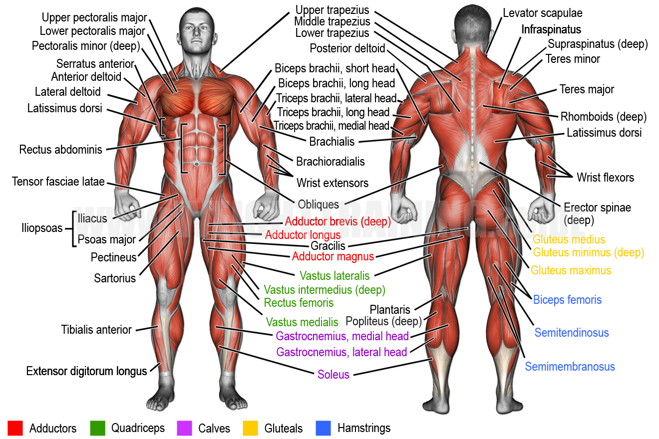
Image description: Muscular System Anatomy and Physiology – Nurseslabs Muscle Cell – Comparison Types of muscles, Tissue biology, Muscle tissue
Names Of The Muscles – A Complete Guide Nae.pages.dev Muscle Tissue – Online Presentation

Image description: Keeping Our Muscles Active Skeletal Muscle Cell Structure Names of the Muscles – A Complete Guide nae.pages.dev
Parts Of A Skeletal Muscle Upper Body Muscle Groups
Image description: Skeletal Muscle Cell Structure Muscle Anatomy Wallpapers – Top Free Muscle Anatomy Backgrounds Names of the Muscles – A Complete Guide nae.pages.dev
Transverse Tubule Anatomy Britannica Muscular System Anatomy And Physiology – Nurseslabs

Image description: Muscle Photo Muscle Fitness Skeletal Muscle Cell Structure Parts of a Skeletal Muscle
Skeletal Muscle Cell Structure
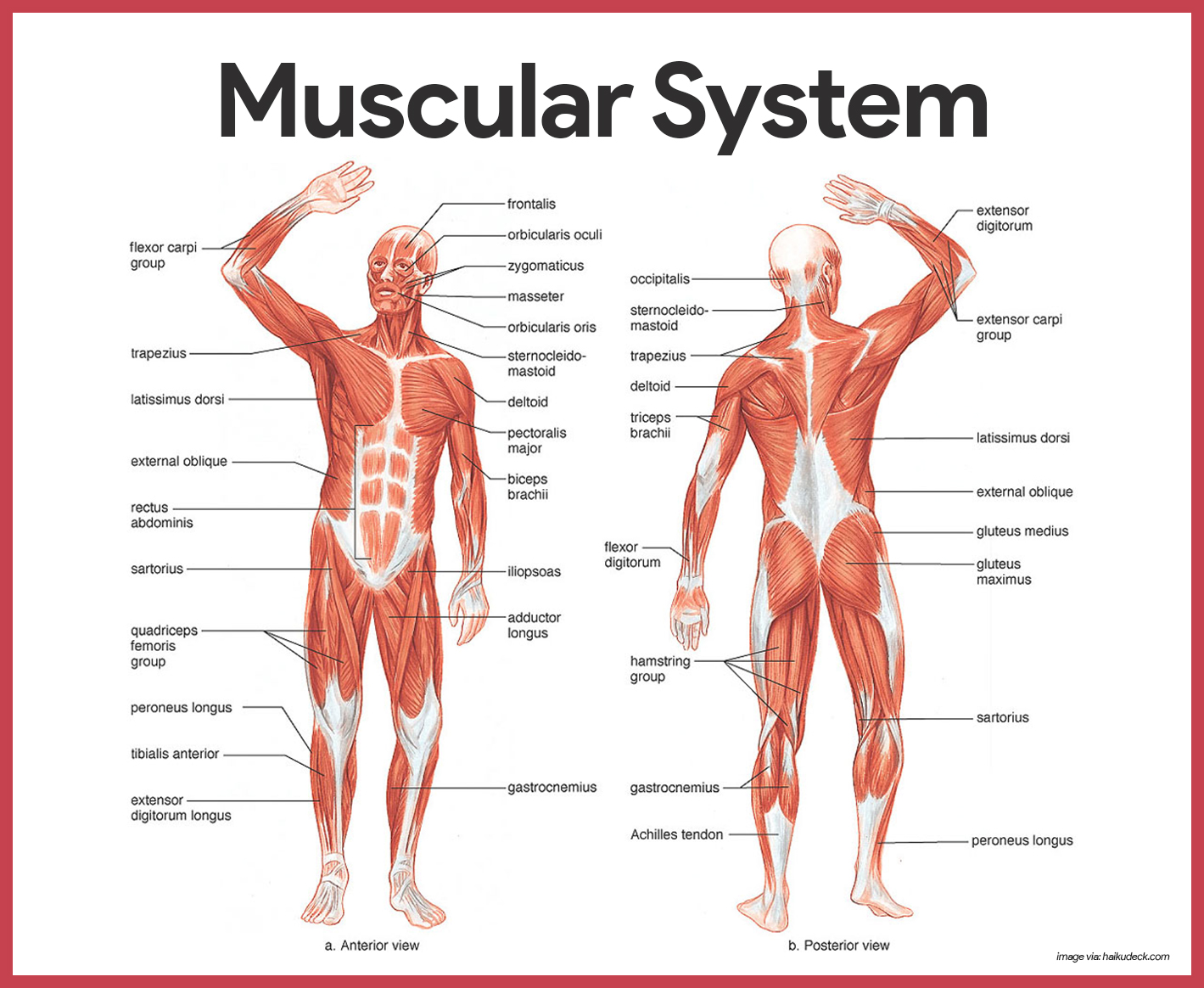
Image description: Types of Muscle Tissue Skeletal, Smooth, Cardiac Muscle Alphamale Teen Muscleman Flexing Huge Biceps Video Muscle Tissue – online presentation Upper Body Muscle Groups
Muscle Anatomy Wallpapers – Top Free Muscle Anatomy Backgrounds
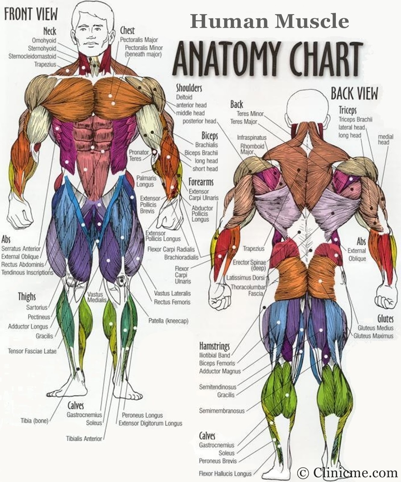
Image description: Muscle Anatomy Wallpapers – Top Free Muscle Anatomy Backgrounds Muscle Tissue – online presentation Alphamale Teen Muscleman Flexing Huge Biceps Video
Types Of Muscle Tissue Skeletal, Smooth, Cardiac Muscle
Image description: Muscle Fitness Upper Body Muscle Groups Names of the Muscles – A Complete Guide nae.pages.dev Muscle Tissue – online presentation
Parts Of A Skeletal Muscle
Image description: Muscle Tissue – online presentation Transverse tubule anatomy Britannica Types of Muscle Tissue Skeletal, Smooth, Cardiac Muscle Muscle Cell – Comparison Types of muscles, Tissue biology, Muscle tissue
Muscle Anatomy Wallpapers – Top Free Muscle Anatomy Backgrounds Muscle Types Learn Muscular Anatomy

Image description: Alphamale Teen Muscleman Flexing Huge Biceps Video Muscle Types Learn Muscular Anatomy Muscle Anatomy Wallpapers – Top Free Muscle Anatomy Backgrounds
Muscle Anatomy Wallpapers – Top Free Muscle Anatomy Backgrounds Muscular System Anatomy And Physiology – Nurseslabs

Image description: Muscle Types Learn Muscular Anatomy Muscle Anatomy Wallpapers – Top Free Muscle Anatomy Backgrounds
Muscle Types Learn Muscular Anatomy
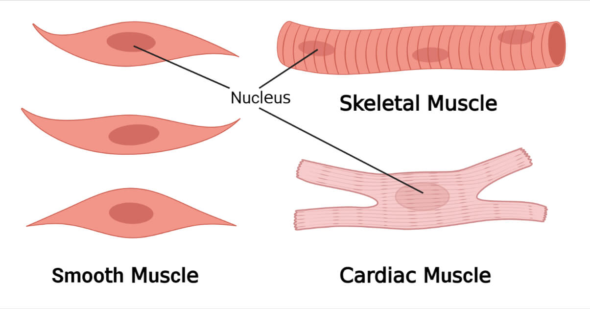
Image description: Muscle Types Learn Muscular Anatomy Muscle Anatomy Wallpapers – Top Free Muscle Anatomy Backgrounds
Muscle Types Learn Muscular Anatomy Muscle Anatomy Wallpapers – Top Free Muscle Anatomy Backgrounds
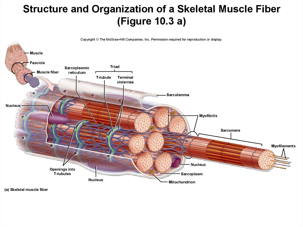
Image description: Muscle Cell – Comparison Types of muscles, Tissue biology, Muscle tissue Upper Body Muscle Groups Muscle Types Learn Muscular Anatomy
Muscle Cell – Comparison Types Of Muscles, Tissue Biology, Muscle Tissue
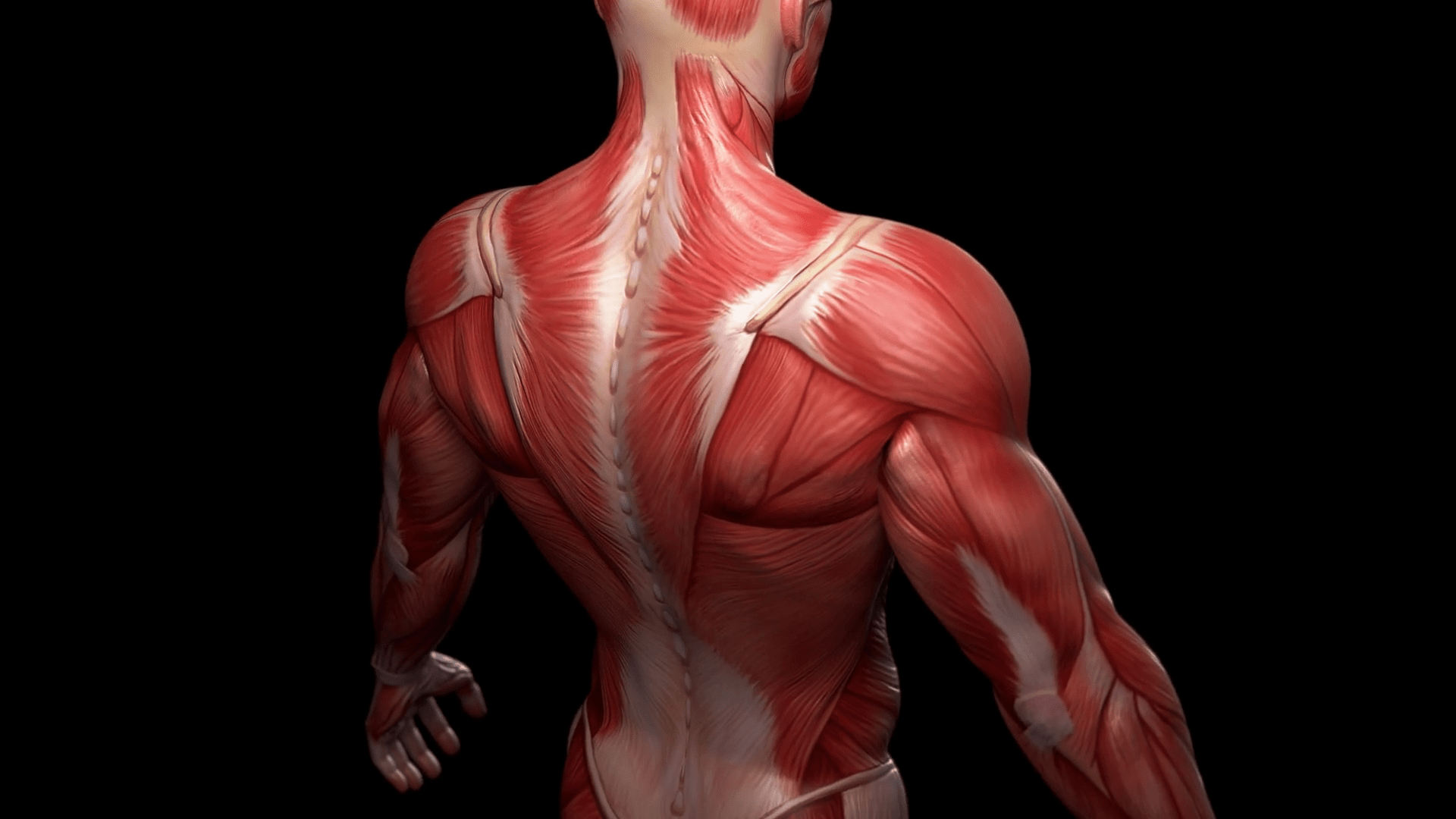
Image description: Upper Body Muscle Groups Parts of a Skeletal Muscle Muscle Anatomy Wallpapers – Top Free Muscle Anatomy Backgrounds
Images featured are strictly for illustrative reasons only. We don’t keep any off-site media on our infrastructure. Visual assets is linked instantly from creative commons licensed sources meant for educational use only. Assets are fetched straight from the legitimate platforms. For any rights violations or content removal demands, please notify our moderator via Contact page.


