

The posterior chest wall, often overlooked, plays a crucial role in respiration, posture, and shoulder movement. Understanding the muscles that make up this area is essential for anyone interested in anatomy, fitness, or rehabilitation. This post will explore the key posterior chest wall muscles, outlining their origins, insertions, actions, and relevant clinical considerations.
The primary muscles residing on the posterior chest wall contribute significantly to upper body stability and movement. Among the most prominent are the **trapezius**, **latissimus dorsi**, and **rhomboids (major and minor)**. Although technically the **serratus posterior superior and inferior** are also located in this region, they are thin, often rudimentary muscles involved in rib elevation and depression and are primarily respiratory muscles.
The **trapezius** is a large, superficial muscle that spans the upper back and neck. It originates from the occipital bone, ligamentum nuchae, and spinous processes of vertebrae C7-T12. Its fibers insert onto the clavicle, acromion, and spine of the scapula. The trapezius performs various actions depending on the section of the muscle engaged. These include elevating, depressing, retracting, and rotating the scapula, as well as extending the head and neck. This broad range of functions makes it vital for shoulder movement and head posture.
The **latissimus dorsi**, often referred to as “lats,” is the broadest muscle in the back. It originates from the spinous processes of vertebrae T7-L5, thoracolumbar fascia, iliac crest, and lower ribs. It inserts into the intertubercular groove of the humerus. The latissimus dorsi is a powerful adductor, internal rotator, and extensor of the arm, playing a key role in activities like swimming, climbing, and rowing. Its connection to the thoracolumbar fascia also contributes to trunk stability.
The **rhomboids (major and minor)** lie deep to the trapezius. The rhomboid minor originates from the spinous processes of vertebrae C7-T1 and the rhomboid major originates from the spinous processes of vertebrae T2-T5. Both insert onto the medial border of the scapula. The rhomboids work together to retract and rotate the scapula, fixing it to the thoracic wall. They also assist in elevating the scapula and stabilizing it during movements of the upper extremity.
Finally, the **serratus posterior superior** and **serratus posterior inferior** muscles, while smaller, also contribute to the posterior chest wall. The superior originates from the spinous processes of C7-T3 and inserts onto ribs 2-5, elevating the ribs. The inferior originates from the spinous processes of T11-L2 and inserts onto ribs 9-12, depressing the ribs. Their actions assist in forced inspiration and expiration, respectively.
Frequently Asked Questions (FAQ)
Q: What happens if the posterior chest wall muscles are weak?
A: Weakness in these muscles can lead to poor posture, shoulder instability, difficulty with overhead activities, and increased risk of injury. It can also contribute to back pain and breathing difficulties.
Q: How can I strengthen my posterior chest wall muscles?
A: Exercises such as rows, pull-ups, lat pulldowns, shrugs, and scapular retractions are effective for strengthening these muscles. Consult a physical therapist or qualified fitness professional for personalized recommendations.
Q: What is scapular winging, and how does it relate to these muscles?
A: Scapular winging is a condition where the medial border of the scapula protrudes away from the rib cage. It can be caused by weakness or paralysis of muscles that stabilize the scapula, such as the serratus anterior (primarily anterior chest wall) and rhomboids.
If you are looking for Anatomical Terms of Location – Anterior – Posterior – TeachMeAnatomy Posterior hip muscles Superficial Diagram Quizlet you’ve visit to the right place. We have 15 Pics about Posterior View of the Trunk Diagram Quizlet, 3 Human Muscle Anatomy Poster Anterior Posterior and Deep – Etsy Australia, Tibialis Posterior Explained Morningside Acupuncture NYC like PPT – The Human Body PowerPoint Presentation, free download – ID3790967 Naming Skeletal Muscles Anatomy and Physiology I. Read more:
PPT – The Human Body PowerPoint Presentation, Free Download – ID3790967
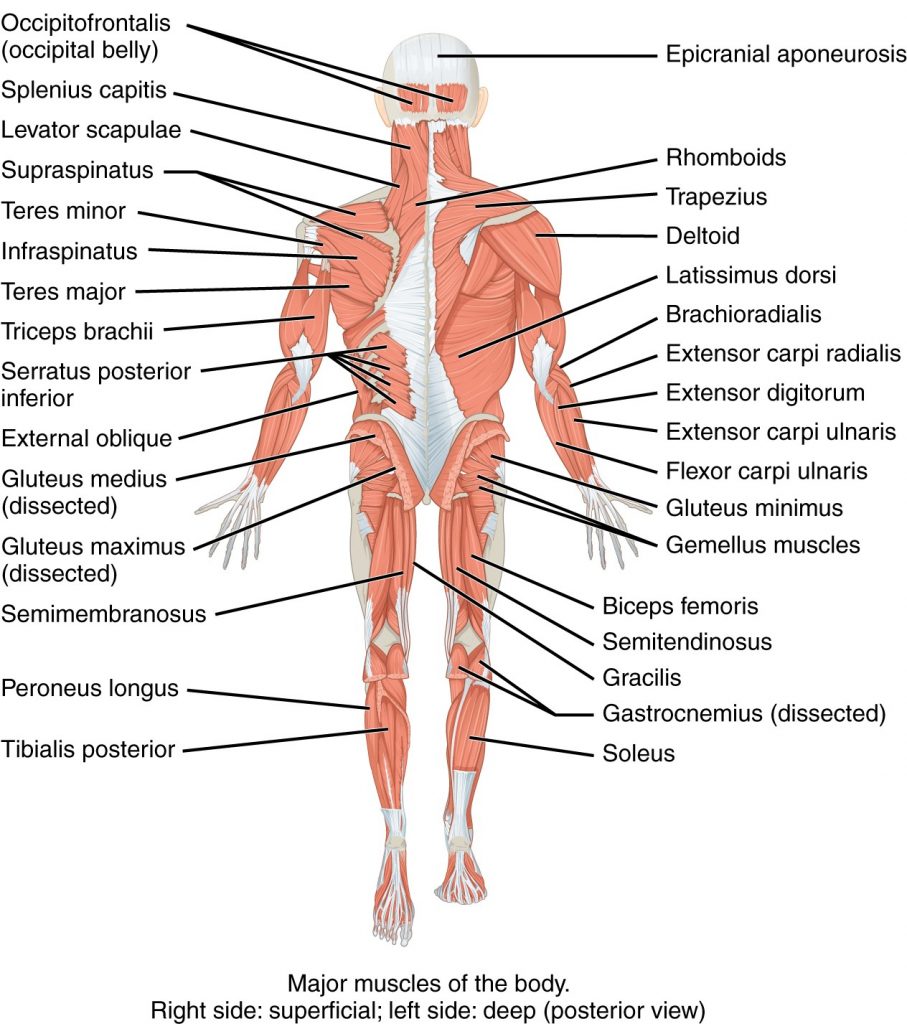
Image description: Posterior Forearm Tattoos Tibialis Posterior Explained Morningside Acupuncture NYC Posterior Skeletal Muscles
Anatomy Directional Terms Anterior Vs Posterior Study Notes, Examples Muscles Of The Posterior Leg – Attachments – Actions – TeachMeAnatomy

Image description: HUMAN BEING ANATOMY MUSCLES POSTERIOR VIEW image – Visual PPT – The Human Body PowerPoint Presentation, free download – ID3790967 Naming Skeletal Muscles Anatomy and Physiology I 3 Human Muscle Anatomy Poster Anterior Posterior and Deep – Etsy Australia
Intermediate Back Muscles AnatomyZone 3 Human Muscle Anatomy Poster Anterior Posterior And Deep – Etsy Australia

Image description: Basic Anatomy Physiology of the Musculoskeletal System Medical Muscles of the Posterior Leg – Attachments – Actions – TeachMeAnatomy 3 Human Muscle Anatomy Poster Anterior Posterior and Deep – Etsy Australia Naming Skeletal Muscles Anatomy and Physiology I
Anatomical Terms Of Location – Anterior – Posterior – TeachMeAnatomy Posterior Anterior Superior Inferior
Image description: Intermediate Back Muscles AnatomyZone Muscles of the Posterior Leg – Attachments – Actions – TeachMeAnatomy Anatomical Terms of Location – Anterior – Posterior – TeachMeAnatomy
Anatomy Directional Terms Anterior Vs Posterior Study Notes, Examples

Image description: Anatomical Terms of Location – Anterior – Posterior – TeachMeAnatomy HUMAN BEING ANATOMY MUSCLES POSTERIOR VIEW image – Visual
Naming Skeletal Muscles Anatomy And Physiology I Posterior View Of The Trunk Diagram Quizlet
Image description: Tibialis Posterior Explained Morningside Acupuncture NYC Muscles of the Posterior Leg – Attachments – Actions – TeachMeAnatomy
Posterior Hip Muscles Superficial Diagram Quizlet Naming Skeletal Muscles Anatomy And Physiology I

Image description: Posterior Forearm Tattoos Basic Anatomy Physiology of the Musculoskeletal System Medical
Posterior Hip Muscles Superficial Diagram Quizlet
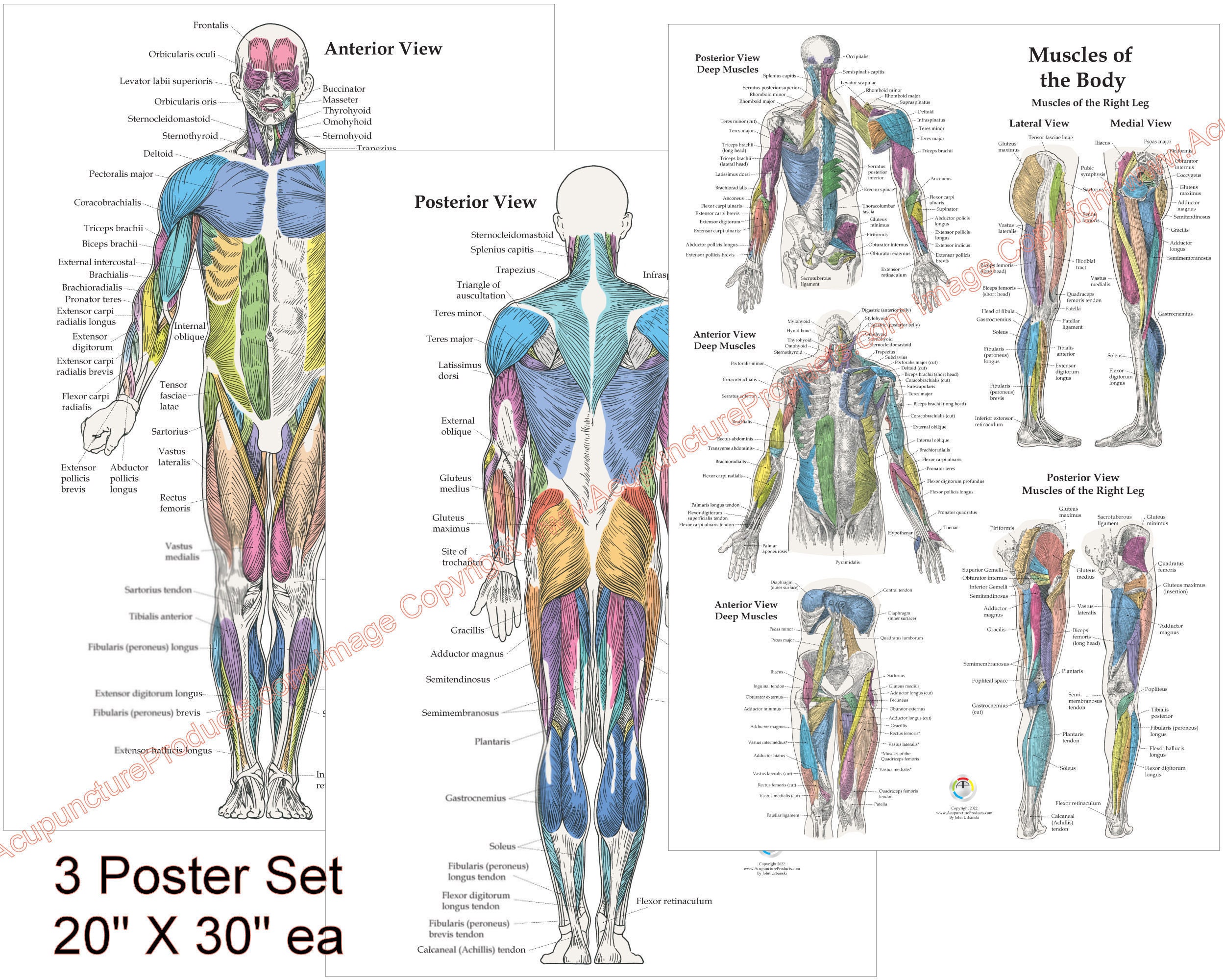
Image description: 3 Human Muscle Anatomy Poster Anterior Posterior and Deep – Etsy Australia Posterior Skeletal Muscles Basic Anatomy Physiology of the Musculoskeletal System Medical Anatomy Directional Terms Anterior vs Posterior Study Notes, Examples
PPT – The Human Body PowerPoint Presentation, Free Download – ID3790967 Naming Skeletal Muscles Anatomy And Physiology I

Image description: Posterior Anterior Superior Inferior 3 Human Muscle Anatomy Poster Anterior Posterior and Deep – Etsy Australia Anatomy Directional Terms Anterior vs Posterior Study Notes, Examples
Posterior Hip Muscles Superficial Diagram Quizlet Naming Skeletal Muscles Anatomy And Physiology I

Image description: Intermediate Back Muscles AnatomyZone Tibialis Posterior Explained Morningside Acupuncture NYC HUMAN BEING ANATOMY MUSCLES POSTERIOR VIEW image – Visual
HUMAN BEING ANATOMY MUSCLES POSTERIOR VIEW Image – Visual

Image description: Tibialis Posterior Explained Morningside Acupuncture NYC Anatomical Terms of Location – Anterior – Posterior – TeachMeAnatomy Posterior View of the Trunk Diagram Quizlet Posterior hip muscles Superficial Diagram Quizlet
Tibialis Posterior Explained Morningside Acupuncture NYC
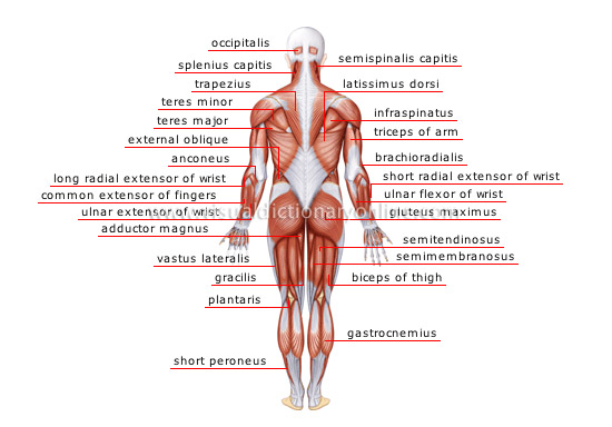
Image description: Intermediate Back Muscles AnatomyZone Basic Anatomy Physiology of the Musculoskeletal System Medical PPT – The Human Body PowerPoint Presentation, free download – ID3790967 Posterior View of the Trunk Diagram Quizlet
Posterior Skeletal Muscles Basic Anatomy Physiology Of The Musculoskeletal System Medical

Image description: Posterior View of the Trunk Diagram Quizlet Basic Anatomy Physiology of the Musculoskeletal System Medical Naming Skeletal Muscles Anatomy and Physiology I PPT – The Human Body PowerPoint Presentation, free download – ID3790967
PPT – The Human Body PowerPoint Presentation, Free Download – ID3790967 Naming Skeletal Muscles Anatomy And Physiology I
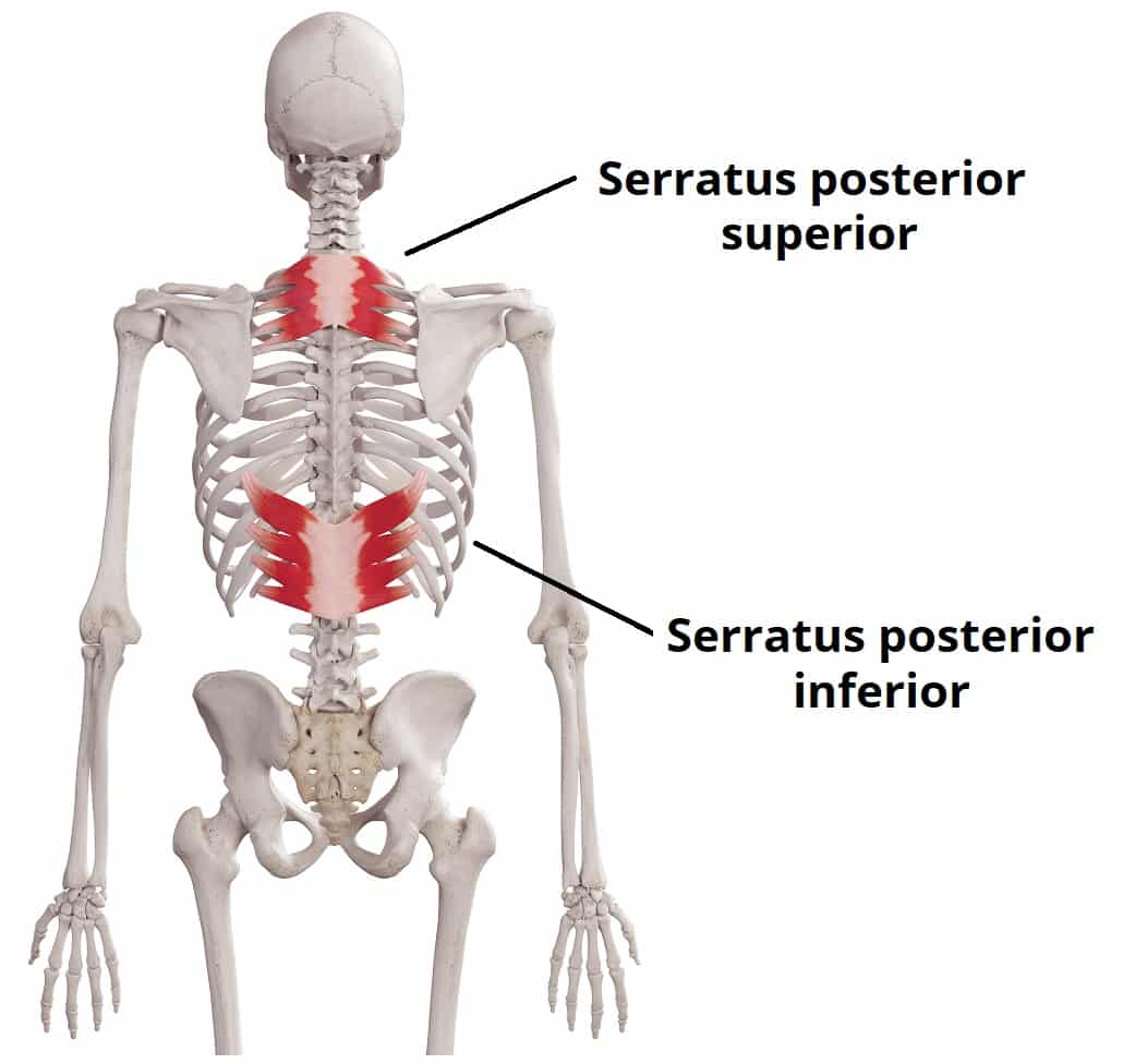
Image description: HUMAN BEING ANATOMY MUSCLES POSTERIOR VIEW image – Visual Posterior hip muscles Superficial Diagram Quizlet Posterior View of the Trunk Diagram Quizlet
3 Human Muscle Anatomy Poster Anterior Posterior And Deep – Etsy Australia

Image description: Posterior hip muscles Superficial Diagram Quizlet Posterior Anterior Superior Inferior Muscles of the Posterior Leg – Attachments – Actions – TeachMeAnatomy Posterior Skeletal Muscles
Images featured are purely for educational purposes only. Our servers do not store any outside media on our system. All resources is linked seamlessly from copyright-free sources shared for informative use only. Downloads are delivered straight from the official platforms. For any ownership inquiries or deletion requests, please contact our site team through our Contact page.


