

The iliopsoas muscle, the most powerful hip flexor in the human body, is a frequent subject of interest in musculoskeletal MRI. Understanding its normal appearance, common pathologies, and relevant imaging findings is crucial for accurate diagnosis and treatment planning. This post provides a concise overview of the iliopsoas muscle on MRI.
On MRI, the iliopsoas muscle is typically evaluated using axial, coronal, and sagittal sequences. Its appearance varies depending on the pulse sequence used. On T1-weighted images, the muscle displays intermediate signal intensity, similar to other skeletal muscles. The borders of the muscle should appear well-defined and smooth. Within the muscle, subtle variations in signal intensity can be seen, reflecting the arrangement of muscle fibers and connective tissue. On T2-weighted images, the normal muscle generally maintains low signal intensity. However, small amounts of fluid within the muscle sheaths or surrounding structures can cause localized areas of increased signal. This is especially important to assess when evaluating for possible pathology. Fat-saturated sequences (e.g., STIR, T2-weighted fat-saturated) are invaluable for detecting fluid, edema, or inflammation within the iliopsoas muscle and adjacent structures.
Several pathologies can affect the iliopsoas muscle, and MRI plays a critical role in their diagnosis. Iliopsoas tendinitis or bursitis often presents with pain and limited range of motion. MRI findings may include thickening of the iliopsoas tendon, increased signal intensity on T2-weighted images (representing edema), and fluid distention within the iliopsoas bursa. Muscle strains or tears can also occur, particularly in athletes. MRI will demonstrate disruption of the muscle fibers, edema, and potentially hematoma formation. Abscesses involving the iliopsoas muscle are relatively rare but serious conditions. MRI will show a fluid collection with surrounding inflammation and potentially involvement of adjacent structures, such as the spine or bowel. Finally, masses, both benign and malignant, can arise within or adjacent to the iliopsoas muscle. MRI is essential for characterizing the lesion, determining its extent, and assessing its relationship to surrounding structures.
Careful interpretation of MRI findings in conjunction with the patient’s clinical presentation is essential for accurate diagnosis and appropriate management of iliopsoas-related conditions. The presence of contrast enhancement, the pattern of edema, and the involvement of adjacent structures are all important factors to consider.
FAQ: Iliopsoas Muscle and MRI
- What is the best MRI sequence to visualize iliopsoas muscle inflammation?
Fat-saturated T2-weighted sequences (like STIR) are highly sensitive for detecting fluid and edema associated with inflammation.
- What is the normal appearance of the iliopsoas muscle on MRI?
It appears with intermediate signal on T1-weighted images and low signal on T2-weighted images. The borders should be well-defined.
- What can cause high signal intensity in the iliopsoas muscle on T2-weighted MRI?
Inflammation, edema, muscle strain or tear, abscess formation, or even a mass can cause increased signal intensity.
- Can MRI differentiate between iliopsoas tendinitis and bursitis?
Yes, MRI can often differentiate between these conditions. Tendinitis typically shows thickening and increased signal within the tendon, while bursitis demonstrates fluid distention within the bursa.
If you are looking for Iliopsoas Bursa Anatomy The Iliopsoasaka the Psoasaka the Eminem Muscle Musculoskeletal Key you’ve came to the right place. We have 15 Images about Iliopsoas Anatomy, Whats Causing Your Snapping Hip Syndrome Clinical Somatics, Iliopsoas Bursa Anatomy like Iliopsoas Bursa Anatomy Iliopsoas The Wonder Muscle That No One Knows. Read more:
Iliopsoas The Wonder Muscle That No One Knows
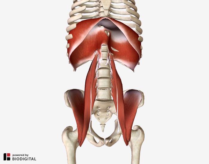
Image description: Iliopsoas Bursitis Stretches human anatomy drawing iliopsoas Stock Photo – Alamy Iliopsoas Complex – Functional Anatomy – Integrative Works
Human Anatomy Drawing Iliopsoas Stock Photo – Alamy Iliopsoas Bursitis Stretches

Image description: Iliopsoas Bursa Anatomy LEONG Orthopaedic Health Iliopsoas Muscle – Personal Training North Iliopsoas The Wonder Muscle That No One Knows
Hip Joint Anatomy Concise Medical Knowledge

Image description: Iliopsoas Bursa Anatomy The Iliopsoasaka the Psoasaka the Eminem Muscle Musculoskeletal Key Iliopsoas The Wonder Muscle That No One Knows Iliopsoas muscles, artwork Artwork, Muscle, Stock images
Iliopsoas – Clinics In Sports Medicine
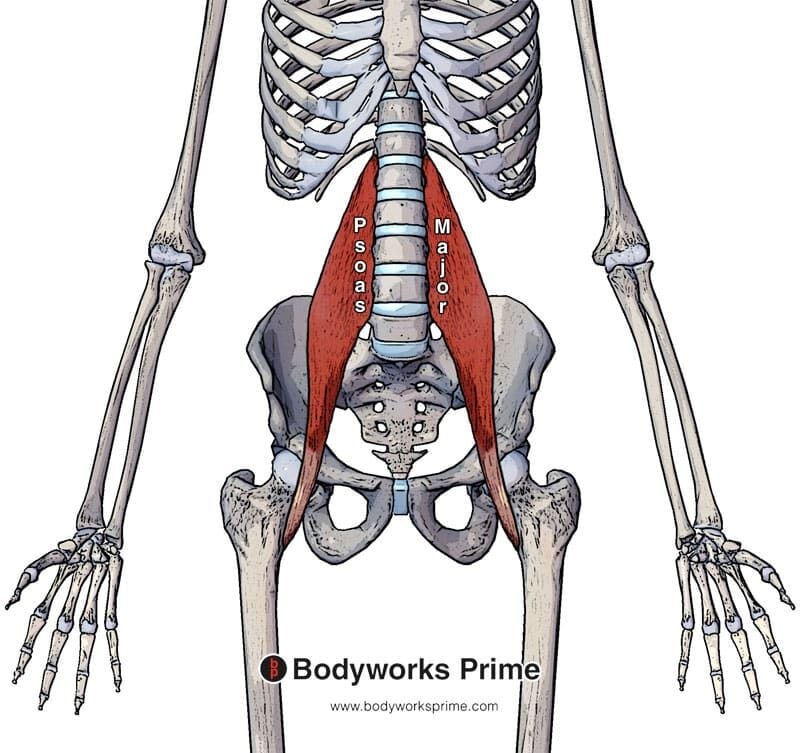
Image description: Human anatomy drawing iliopsoas Stock Photo – Alamy The Iliopsoasaka the Psoasaka the Eminem Muscle Musculoskeletal Key Iliopsoas The Wonder Muscle That No One Knows Hip Joint Anatomy Concise Medical Knowledge
Iliopsoas – Clinics In Sports Medicine Iliopsoas Bursa Anatomy

Image description: LEONG Orthopaedic Health Iliopsoas Muscle – Personal Training North human anatomy drawing iliopsoas Stock Photo – Alamy The Iliopsoasaka the Psoasaka the Eminem Muscle Musculoskeletal Key Psoas Iliacus muscles Yoga anatomy, Muscle, Energy healing
Iliopsoas Anatomy

Image description: Iliopsoas Anatomy Iliopsoas Bursitis Stretches Psoas Iliacus muscles Yoga anatomy, Muscle, Energy healing
Iliopsoas Anatomy Iliopsoas – Clinics In Sports Medicine
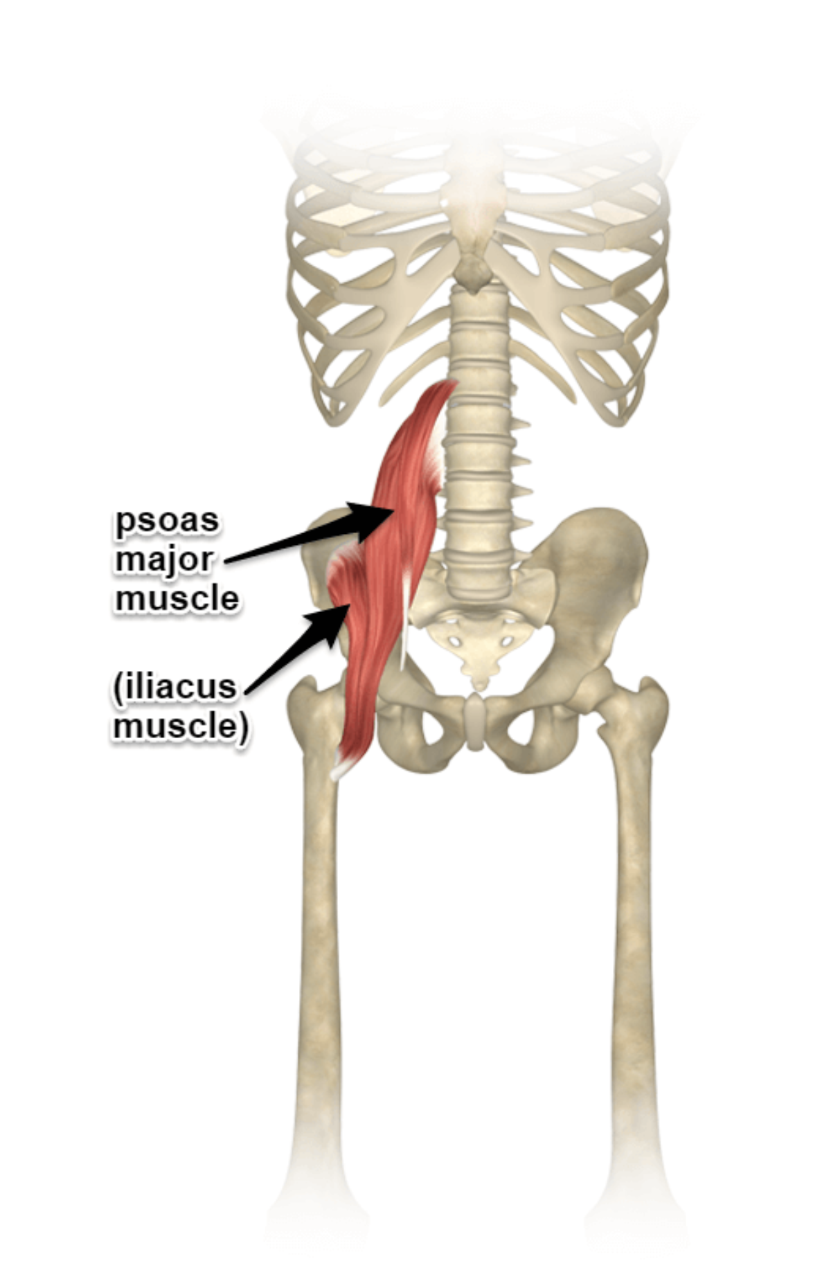
Image description: Iliopsoas Bursa Anatomy Iliopsoas – Clinics in Sports Medicine LEONG Orthopaedic Health Iliopsoas Muscle – Personal Training North Iliopsoas Bursitis Stretches
Human Anatomy Drawing Iliopsoas Stock Photo – Alamy Iliopsoas Bursitis Stretches
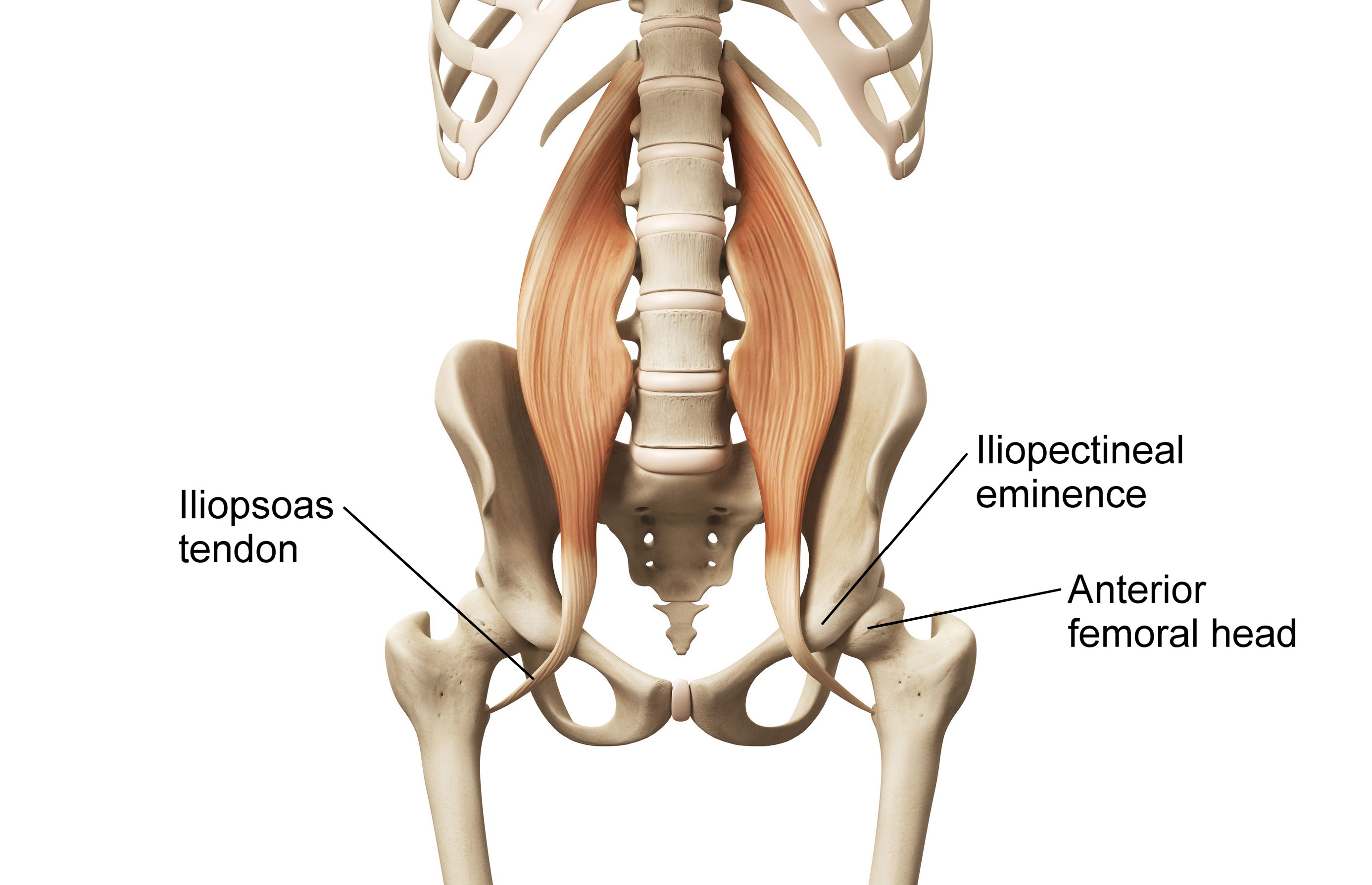
Image description: Iliopsoas – Clinics in Sports Medicine LEONG Orthopaedic Health Iliopsoas Muscle – Personal Training North Whats Causing Your Snapping Hip Syndrome Clinical Somatics
Iliopsoas The Wonder Muscle That No One Knows

Image description: Human anatomy drawing iliopsoas Stock Photo – Alamy The Iliopsoasaka the Psoasaka the Eminem Muscle Musculoskeletal Key Iliopsoas Bursa Anatomy
Iliopsoas Bursa Anatomy LEONG Orthopaedic Health Iliopsoas Muscle – Personal Training North

Image description: LEONG Orthopaedic Health Iliopsoas Muscle – Personal Training North human anatomy drawing iliopsoas Stock Photo – Alamy Iliopsoas Anatomy
Iliopsoas Muscles, Artwork Artwork, Muscle, Stock Images Iliopsoas Anatomy
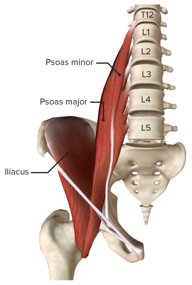
Image description: Hip Joint Anatomy Concise Medical Knowledge LEONG Orthopaedic Health Iliopsoas Muscle – Personal Training North Iliopsoas muscles, artwork Artwork, Muscle, Stock images Iliopsoas Anatomy
LEONG Orthopaedic Health Iliopsoas Muscle – Personal Training North
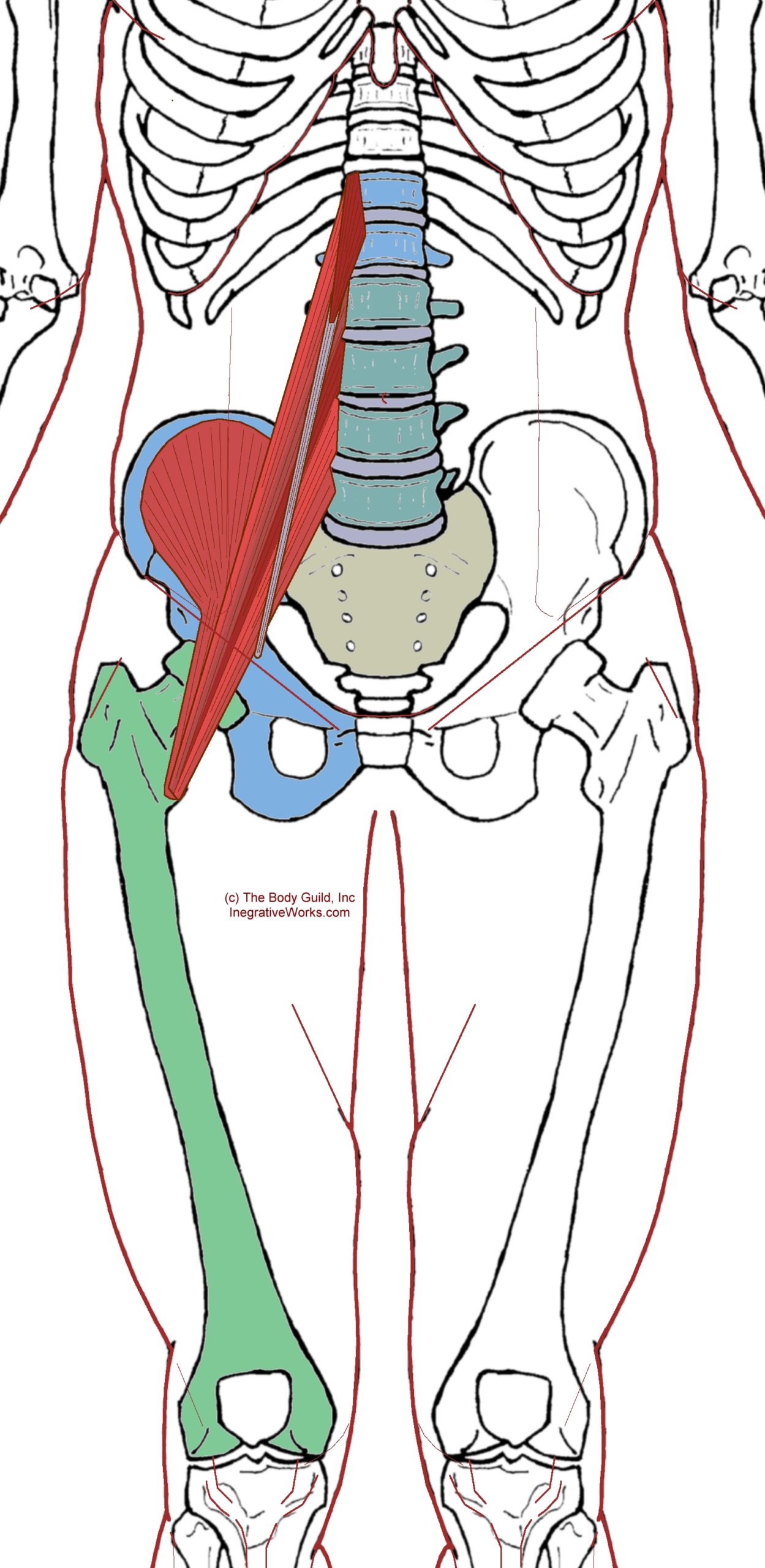
Image description: Iliopsoas Anatomy Iliopsoas The Wonder Muscle That No One Knows Iliopsoas Bursa Anatomy Psoas Iliacus muscles Yoga anatomy, Muscle, Energy healing
The Iliopsoasaka The Psoasaka The Eminem Muscle Musculoskeletal Key Iliopsoas Bursitis Stretches

Image description: Iliopsoas Bursitis Stretches Iliopsoas Anatomy The Iliopsoasaka the Psoasaka the Eminem Muscle Musculoskeletal Key
Hip Joint Anatomy Concise Medical Knowledge Whats Causing Your Snapping Hip Syndrome Clinical Somatics

Image description: Whats Causing Your Snapping Hip Syndrome Clinical Somatics LEONG Orthopaedic Health Iliopsoas Muscle – Personal Training North Iliopsoas Anatomy Iliopsoas Bursa Anatomy
Whats Causing Your Snapping Hip Syndrome Clinical Somatics

Image description: The Iliopsoasaka the Psoasaka the Eminem Muscle Musculoskeletal Key Psoas Iliacus muscles Yoga anatomy, Muscle, Energy healing Hip Joint Anatomy Concise Medical Knowledge Iliopsoas Bursa Anatomy
Illustrations provided are entirely for explanatory objectives only. We refrain from hosting any third-party media on our servers. All resources is streamed automatically from creative commons licensed sources meant for informative use only. Downloads are delivered straight from the source distributors. For any intellectual property issues or content removal demands, please get in touch with our site team through Contact page.


